
Fascicular block
Left posterior fascicular block
The rear division of the left bundle is blocked, so the left ventricle is activated from above downward and the axis swings right. Rare in isolation.
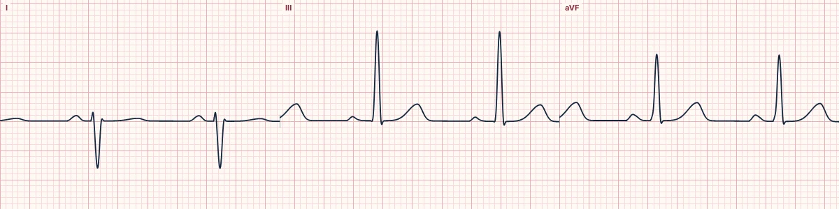
What the heart is doing
The impulse reaches the left ventricle only through the anterior fascicle. The upper lateral wall is activated first and the wave spreads downward and rightward, pulling the axis right.
Why it looks this way
This is LPFB because the axis is beyond +90° with rS complexes in I and aVL, qR complexes in III and aVF and a narrow QRS.
Why it matters
Uncommon alone because the posterior fascicle is broad with a dual blood supply; its presence implies more extensive conduction disease. With RBBB it forms a higher-risk bifascicular block.
What to look for
- Lead I is predominantly negative while III and aVF are strongly positive.
- The QRS remains narrow.
- Rule out other causes of right axis deviation first; this is a diagnosis of exclusion.
Frequently asked questions
What are the ECG criteria for left posterior fascicular block?
Axis: +90° to +180°. QRS: < 120 ms. I and aVL: small r, deep S (rS). III and aVF: small q, tall R (qR). Exclusion: no right ventricular hypertrophy or lateral infarct.
How is left posterior fascicular block identified on an ECG?
This is LPFB because the axis is beyond +90° with rS complexes in I and aVL, qR complexes in III and aVF and a narrow QRS.
What can left posterior fascicular block be mistaken for?
Right ventricular hypertrophy: tall R in V1 and right atrial enlargement. Acute pulmonary embolism: sinus tachycardia with new right axis shift. Arm lead reversal: P wave and T wave also inverted in lead I.
What causes left posterior fascicular block?
Common causes: coronary artery disease, inferior infarction; cardiomyopathy; degenerative conduction disease; myocarditis; and hypertension.