
Position & lead placement
Arm lead reversal (LA/RA)
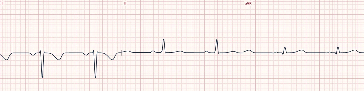
The left-arm and right-arm electrodes have been swapped. Lead I is flipped upside down and aVR and aVL trade places, while the chest leads are unaffected.
What the heart is doing
Lead I measures left arm minus right arm, so swapping the electrodes reverses its polarity. The leads built from each arm exchange identities, while the chest electrodes are untouched.
Why it looks this way
This is arm lead reversal because lead I is completely inverted and aVR is upright while the precordial leads show normal R-wave progression.
Why it matters
The most common recording error. It carries no risk to the patient except misdiagnosis: it imitates lateral infarction, an ectopic atrial rhythm or dextrocardia. Always repeat the recording.
What to look for
- An upright P wave and QRS in aVR is almost always a lead error.
- Lead I and V6 normally look alike; if I is inverted and V6 is normal, the arm leads are swapped.
- Check the electrodes and repeat the ECG before interpreting anything else.
Frequently asked questions
What are the ECG criteria for arm lead reversal (LA/RA)?
Lead I: P wave, QRS and T wave all inverted. aVR and aVL: swapped: aVR looks upright. II and III: swapped. aVF: unchanged. V1–V6: normal R-wave progression.
How is arm lead reversal (LA/RA) identified on an ECG?
This is arm lead reversal because lead I is completely inverted and aVR is upright while the precordial leads show normal R-wave progression.
What can arm lead reversal (LA/RA) be mistaken for?
Dextrocardia: the R wave also shrinks from V1 to V6. Ectopic atrial rhythm: P wave inverted but QRS and T upright in lead I. High lateral infarction: Q waves in I and aVL with an upright P wave.
What causes arm lead reversal (LA/RA)?
Common causes: electrodes attached to the wrong arms; cables plugged into the wrong electrodes; tangled or unlabeled lead wires; and hurried acquisition in an emergency.