
Age & normal variants
Draft — pending owner review
Normal infant ECG pattern
Right ventricular dominance and juvenile T waves
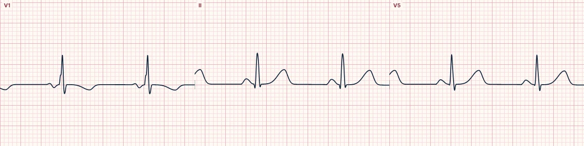
A normal ECG for an infant: fast rate, rightward axis, tall R waves in V1 and inverted T waves in V1–V3. In an adult these would be abnormal; in a baby they are expected.
What the heart is doing
Before birth the right ventricle pumps against high pulmonary resistance and is as thick as the left. Its forces dominate the infant ECG, pointing the QRS rightward and anterior, until the left ventricle grows to dominate.
Why it looks this way
This is the normal infant ECG pattern because a fast sinus rhythm has a rightward axis, a dominant R wave in V1 and inverted T waves in V1–V3.
Why it matters
Adult criteria misread normal children: a tall R in V1 is not right ventricular hypertrophy in an infant, and inverted T waves in V1–V3 are expected. Use age-specific tables, and ask for pediatric cardiology review when unsure.
What to look for
- Check the age before reading anything: normal values change through childhood.
- An upright T wave in V1 from one week to adolescence can mean right ventricular hypertrophy.
- Compare R and S in V1 and V6 against age-specific normal tables.
Frequently asked questions
What are the ECG criteria for normal infant ECG pattern?
Rate: about 100–160/min in infancy. Axis: rightward: about +30° to +180° at birth, nearer +10° to +120° by one year. V1: dominant R wave (R larger than S). T waves: inverted in V1–V3 after the first week (juvenile pattern). Intervals: short PR and narrow QRS for age.
How is normal infant ECG pattern identified on an ECG?
This is the normal infant ECG pattern because a fast sinus rhythm has a rightward axis, a dominant R wave in V1 and inverted T waves in V1–V3.
What can normal infant ECG pattern be mistaken for?
Right ventricular hypertrophy: in an adult, or an upright T wave in V1 in a child. Arrhythmogenic right ventricular cardiomyopathy: T inversion in V1–V3 after adolescence. Wellens pattern, type B: deep T inversion in V2–V4 in an adult with chest pain.
What causes normal infant ECG pattern?
Common causes: normal fetal circulation leaves the right ventricle thick at birth; the right ventricle thins over the first years; left ventricular dominance develops through childhood; and juvenile T waves usually resolve by adolescence.