Lessons you can try
Every concept opens in the lab with the right rhythm already loaded, so reading and doing happen together.
 Mastering Cardiology
/
Open the lab
Electrocardiography, taught from the cell up
Watch the impulse leave the sinus node, cross the heart and draw the tracing in front of you. Then change one thing and see what happens.
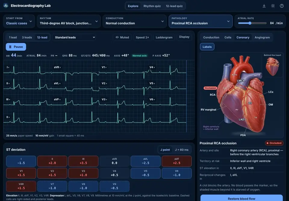
Build any rhythm, add a bundle branch block or an occlusion, and study it on 1, 3 or 12 leads with the heart beating beside it.
Open the lab GrowingAn atlas of electrocardiography. Every entry has its criteria, what to look for, mimics, and a live example one tap away.
Browse the atlas FreeSeven stages from the normal ECG to coronary occlusions, in the order that makes each pattern easy. Your progress is saved as you go.
Start learning Free previewMastering Electrocardiography: the rhythm interpretation textbook and its SAVE method workbook, with 25 pages of each to read here.
See the booksOne heartbeat, slowed down
The heart's own pacemaker sets off the beat. The impulse spreads across both atria, and their thin muscle writes a small, rounded wave.
The only electrical path to the ventricles conducts slowly on purpose, so the atria can finish filling them. Too little tissue is active to move the tracing, and it rests on the baseline.
The impulse races down the bundle of His, the bundle branches and the Purkinje fibres, reaching nearly all of the ventricular muscle in under a tenth of a second. A large muscle activated quickly writes a tall, narrow complex.
Every ventricular cell is depolarized and holding there while calcium enters and the muscle contracts. With no voltage difference across the heart, the tracing is flat again.
The cells repolarize, more slowly and less evenly than they fired, which writes a broad wave. The heart is ready for the next beat.
The Electrocardiography Lab
Most ECG teaching shows you a strip and tells you its name. The lab shows you the heart that made it.
AI ECG Interpreter
Upload a 12-lead or a rhythm strip as a photo, scan or PDF. It is straightened and stripped of patient details on your device, then read the way the lab teaches: rate, rhythm, axis, intervals, lead-by-lead ST and T, occlusion criteria and a differential, with what to check yourself.
No card needed for the free uses. Plans renew monthly and can be cancelled at any time.
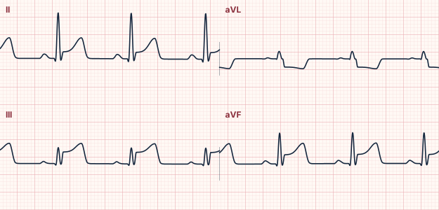
Inferior, probably with posterior extension (right coronary or dominant circumflex)
Books by Adam Thompson, EMT-P, MS
A rhythm interpretation textbook and its companion workbook, built on one routine for every strip, SAVE: speed and regularity, atrial activity, ventricular activity, extra beats. Read 25 pages of each right here before you buy.
The workbook · Illustrated Review Edition
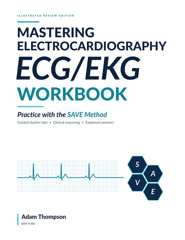
Practice with the SAVE Method. A 40-question baseline check, twelve lessons and 70 ECG cases, with explained answers.
The textbook · Second Edition
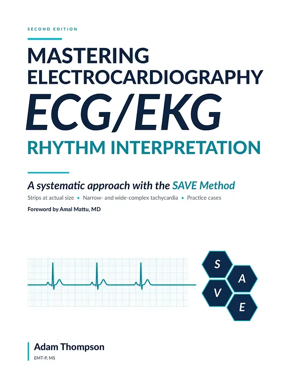
A systematic approach with the SAVE Method. Nineteen chapters with strips at actual size, practice cases and a foreword by Amal Mattu, MD.
Courses
Each course will pair short lessons with hands-on work in the lab, check your understanding as you go and keep your place between visits. None is open yet. Until the first one is, the free learning path shows how it will feel.
Every concept opens in the lab with the right rhythm already loaded, so reading and doing happen together.
Question sets drawn fresh each time: the same diagnosis on a different patient, so you learn the pattern and not the picture.
An account that remembers where you stopped, what you have mastered and what deserves another look.
Coming soon
One email when the first course is ready, and the occasional note about new interpreter features and the books if you choose them. No spam, and you can leave with one click.