
Other conditions
Acute pericarditis
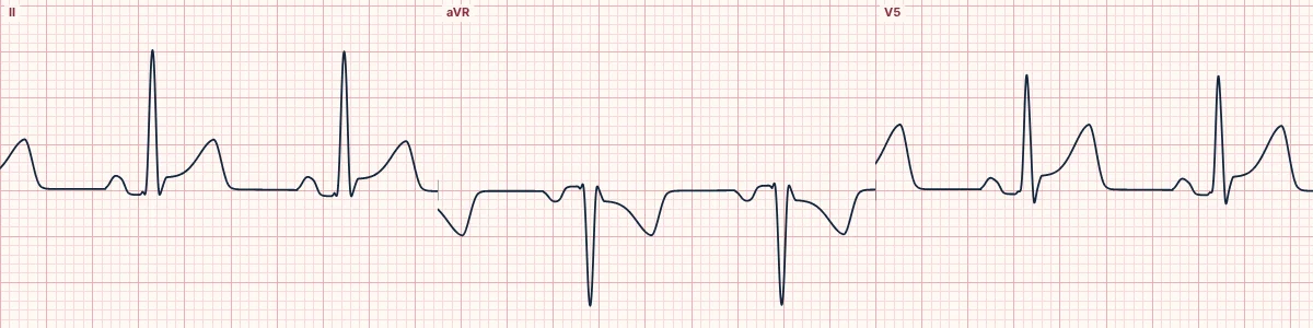
Inflammation of the sac around the heart irritates the muscle surface everywhere, producing widespread concave ST elevation and PR depression.
What the heart is doing
Inflammation injures the outer layer of muscle over the whole heart. The ventricular injury current raises the ST diffusely; a matching atrial injury current displaces the PR segment.
Why it looks this way
This is acute pericarditis because concave ST elevation and PR depression are widespread, with the reverse in aVR and no other reciprocal changes.
Why it matters
Usually self-limiting and treated with anti-inflammatory drugs. Exclude STEMI first, and watch for a pericardial effusion progressing to tamponade or for associated myocarditis.
What to look for
- ST elevation that crosses coronary territories and does not fit one artery.
- PR segments that sit below the baseline, with the opposite in aVR.
- No ST depression in aVL or the inferior leads; if present, think infarction.
Frequently asked questions
What are the ECG criteria for acute pericarditis?
ST segment: concave elevation in most limb and chest leads. PR segment: depressed in the same leads. aVR: ST depression with PR elevation. Reciprocal changes: none apart from aVR (and sometimes V1). Pattern: ST elevation greater in II than in III.
How is acute pericarditis identified on an ECG?
This is acute pericarditis because concave ST elevation and PR depression are widespread, with the reverse in aVR and no other reciprocal changes.
What can acute pericarditis be mistaken for?
STEMI: convex elevation in one territory with reciprocal depression. Early repolarization: no PR depression; ST small relative to the T wave. Inferior STEMI: elevation greater in III than II with depression in aVL.
What causes acute pericarditis?
Common causes: viral or idiopathic; after myocardial infarction or cardiac surgery; kidney failure (uremia); autoimmune disease; cancer; and bacterial or tuberculous infection.