
Repolarization & channelopathies
Benign early repolarization
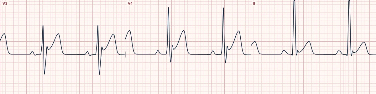
A common normal variant in healthy young people: on a narrow, normally conducted QRS, the ST segment takes off slightly above baseline with a notch at the J point and a tall upright T wave.
What the heart is doing
Differences in early repolarization currents between the outer and inner layers of the ventricular wall create a small voltage gradient at the J point, lifting the start of the ST segment.
Why it looks this way
This is benign early repolarization because concave ST elevation with a notched J point and tall T waves appears without reciprocal ST depression.
Why it matters
Harmless in most people. The danger is missing a subtle LAD occlusion: terminal QRS distortion, Q waves, reciprocal depression, hyperacute T waves or a formula value of 18.2 or more point to occlusion. It is diagnosed only on a narrow QRS; with a bundle branch block, pacing or LVH the ST follows the QRS.
What to look for
- A small notch or hook where the QRS meets the ST segment, best in V4–V6.
- The ST is concave, like a smile, and the T wave is tall but in proportion to a tall QRS.
- V2 and V3 keep an S wave or J wave, with no Q waves in V2–V4 and no reciprocal depression outside aVR.
- Dr. Stephen W. Smith's formula: 0.052·QTc − 0.151·QRS V2 − 0.268·R V4 + 1.062·STE60 V3, in ms and mm.
Frequently asked questions
What are the ECG criteria for benign early repolarization?
QRS: narrow, normally conducted, often high voltage. ST segment: concave elevation, usually < 3 mm; no reciprocal depression except in aVR. J point: notched or slurred ('fish hook'), best in V4–V6. V2 and V3: S wave or J wave present; no Q waves in V2–V4. T waves and QTc: T in proportion to the QRS; QTc normal or short. Smith formula: below 18.2 favours early repolarization; 18.2 or more suggests LAD occlusion.
How is benign early repolarization identified on an ECG?
This is benign early repolarization because concave ST elevation with a notched J point and tall T waves appears without reciprocal ST depression.
What can benign early repolarization be mistaken for?
Anterior STEMI: convex or straight ST, reciprocal depression, changes over minutes. Pericarditis: PR depression and ST elevation large relative to the T wave. Brugada pattern: coved elevation with an inverted T wave in V1–V2.
What causes benign early repolarization?
Common causes: normal variant; young age; athletic conditioning; high vagal tone; and more common in men.