
Repolarization & channelopathies
Draft — pending owner review
Brugada pattern, type 2 (saddleback)
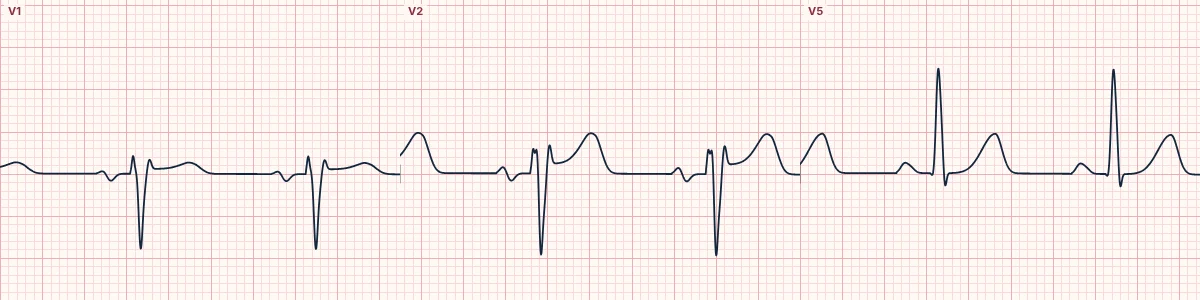
The saddleback Brugada pattern: a high J point in V1–V2, an ST segment that dips but stays raised, then an upright T wave. It is suggestive only; Brugada syndrome needs the type 1 pattern.
What the heart is doing
A milder imbalance between inward sodium and outward potassium currents in the right ventricular outflow epicardium raises the J point without reversing repolarization, so the T wave stays upright.
Why it looks this way
This is a type 2 Brugada pattern because V2 shows a high J-point take-off, a raised ST trough and an upright T wave in a saddleback shape.
Why it matters
A type 2 pattern alone does not establish Brugada syndrome. It calls for a careful history (syncope, nocturnal agonal breathing, family sudden death), repeat ECGs with high leads and expert referral. Treat fever promptly.
What to look for
- Trace V2: a high take-off, a dip, then an upright T wave, like a saddle.
- Look for an r′ in V1–V2 without a wide S wave in I and V6.
- Record V1–V2 one and two rib spaces higher: a type 1 pattern may appear.
Frequently asked questions
What are the ECG criteria for brugada pattern, type 2 (saddleback)?
V1–V2: saddleback ST elevation. J point: high take-off, usually ≥ 2 mm. ST trough: stays ≥ 0.5 mm above the baseline. T wave: upright in V2, variable in V1. Diagnosis: needs conversion to type 1, spontaneously, with fever or on drug challenge.
How is brugada pattern, type 2 (saddleback) identified on an ECG?
This is a type 2 Brugada pattern because V2 shows a high J-point take-off, a raised ST trough and an upright T wave in a saddleback shape.
What can brugada pattern, type 2 (saddleback) be mistaken for?
Brugada pattern: type 1 is coved, sloping into an inverted T wave. Incomplete right bundle branch block: a narrow r′ with no ST elevation. Athlete's heart: benign J-point elevation, usually most marked in V2–V4.
What causes brugada pattern, type 2 (saddleback)?
Common causes: inherited sodium-channel variants; fever; sodium-channel blocking drugs; athletic or normal variant; and V1–V2 placed too high.