
Electrolytes
Hyperkalemia, severe (sine wave)
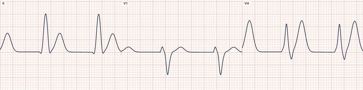
Critically high potassium: P waves vanish and the very wide QRS merges with the T wave into a smooth sine-wave shape. Cardiac arrest is imminent.
What the heart is doing
Severe depolarization of resting cells nearly abolishes sodium-channel function. Atrial muscle stops responding, and ventricular conduction is so slow that depolarization overlaps with repolarization.
Why it looks this way
This is severe hyperkalemia because P waves are absent and the very wide QRS merges with the T wave into a sine-wave pattern.
Why it matters
A pre-arrest rhythm that can turn into VF, pulseless electrical activity or asystole within minutes. Treat on the ECG without waiting for laboratory results: calcium first, then measures to lower potassium.
What to look for
- Broad, rounded, rolling waves in which QRS and T cannot be separated.
- No P waves anywhere.
- A wide, slow, bizarre rhythm in a dialysis patient is hyperkalemia until proven otherwise.
Frequently asked questions
What are the ECG criteria for hyperkalemia, severe (sine wave)?
P waves: absent. QRS: very wide (often > 160 ms), blending into the T wave. Pattern: smooth sine wave with no distinct ST segment. Rate: often slow.
How is hyperkalemia, severe (sine wave) identified on an ECG?
This is severe hyperkalemia because P waves are absent and the very wide QRS merges with the T wave into a sine-wave pattern.
What can hyperkalemia, severe (sine wave) be mistaken for?
Ventricular tachycardia: usually faster, with a distinct QRS. Idioventricular rhythm: wide and slow but with a separate ST–T. Sodium-channel blocker toxicity: wide QRS with a terminal R in aVR.
What causes hyperkalemia, severe (sine wave)?
Common causes: kidney failure or missed dialysis; ACE inhibitors, potassium-sparing diuretics; acidosis, including diabetic ketoacidosis; tissue breakdown (crush injury, rhabdomyolysis, burns); and adrenal insufficiency.