
Ventricular
Idioventricular rhythm
A slow escape rhythm from the ventricles at 20–40/min, appearing when all higher pacemakers have failed. Wide QRS, no P waves.
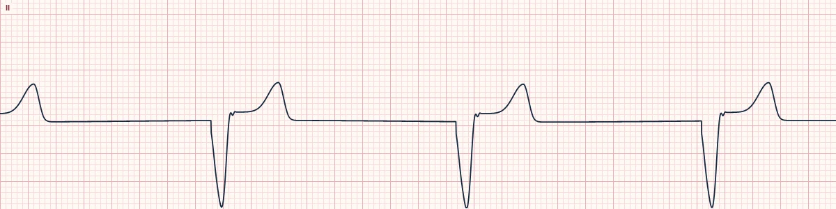
What the heart is doing
With no impulse from the sinus node or AV junction, a ventricular focus fires at its slow intrinsic rate. Activation spreads muscle to muscle, producing a wide QRS.
Why it looks this way
This is an idioventricular rhythm because it is regular at 20–40/min with wide QRS complexes and no P waves.
Why it matters
The heart's last backup pacemaker. Never suppress it with antiarrhythmics, which could cause asystole. If there is a pulse, support the rate and prepare for pacing; if not, treat as cardiac arrest.
What to look for
- Very slow, wide, regular complexes with large T waves pointing the opposite way.
- No P waves anywhere on the strip.
- Check for a pulse immediately; this rhythm is often pulseless.
Frequently asked questions
What are the ECG criteria for idioventricular rhythm?
Rate: 20–40/min. Rhythm: regular. P waves: absent. PR interval: none. QRS: wide (≥ 120 ms), bizarre.
How is idioventricular rhythm identified on an ECG?
This is an idioventricular rhythm because it is regular at 20–40/min with wide QRS complexes and no P waves.
What can idioventricular rhythm be mistaken for?
Third-degree AV block with ventricular escape: P waves are present but unrelated. Junctional escape with bundle branch block: rate 40–60/min. Agonal rhythm: slower, irregular, with deteriorating complexes.
What causes idioventricular rhythm?
Common causes: failure of the sinus node and junction; complete AV block; post-arrest and dying heart; severe hypoxia or acidosis; hyperkalemia; and drug toxicity.