
Ventricular
Ventricular bigeminy
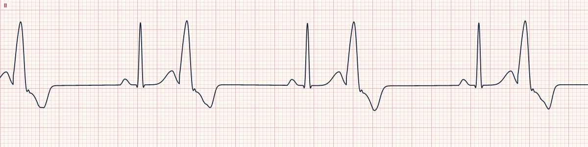
Every normal beat is followed by a PVC, giving a repeating normal–PVC–pause pattern.
What the heart is doing
After each sinus beat, a ventricular focus fires at a fixed interval, often by re-entry in nearby tissue. The pause that follows lets the next sinus beat conduct, and the sequence repeats.
Why it looks this way
This is ventricular bigeminy because every second beat is an early, wide PVC coupled at a fixed interval to the sinus beat before it.
Why it matters
It may cut effective cardiac output if the PVCs do not generate a pulse. Look for and correct ischemia, hypoxia, electrolyte loss or drug toxicity; a high long-term PVC burden can weaken the ventricle.
What to look for
- Pairs of beats: a narrow one then a wide one, followed by a pause.
- The interval from each sinus beat to its PVC is constant.
- Check a pulse. PVCs may not perfuse, so the pulse can be half the monitor rate.
Frequently asked questions
What are the ECG criteria for ventricular bigeminy?
Rate: depends on sinus rate; beats come in pairs. Rhythm: regularly irregular. P waves: before the sinus beats only. QRS: alternating narrow sinus beat and wide PVC. Coupling: fixed interval from sinus beat to PVC.
How is ventricular bigeminy identified on an ECG?
This is ventricular bigeminy because every second beat is an early, wide PVC coupled at a fixed interval to the sinus beat before it.
What can ventricular bigeminy be mistaken for?
Atrial bigeminy: the early beat is narrow and has a P wave. Atrial bigeminy with aberrancy: wide early beat, but preceded by an early P wave. 3:2 Wenckebach: all beats narrow, with a lengthening PR.
What causes ventricular bigeminy?
Common causes: myocardial ischemia; hypokalemia, hypomagnesemia; digoxin toxicity; cardiomyopathy and heart failure; stimulants and caffeine; and hypoxia.