
Ventricular
Accelerated idioventricular rhythm
A ventricular focus firing at 40–110/min, faster than a normal escape but slower than VT. Classically appears when a blocked coronary artery reopens.
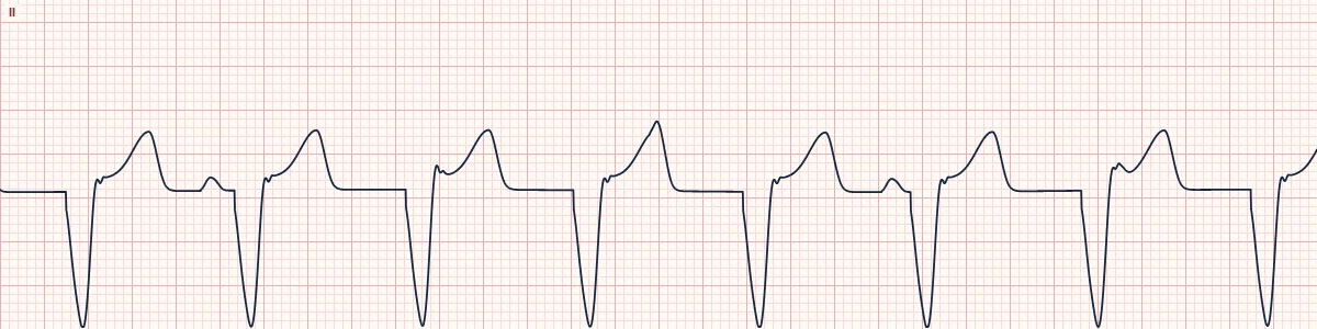
What the heart is doing
Enhanced automaticity in a ventricular focus makes it fire slightly faster than the sinus node, so it takes over. The atria continue under sinus control, dissociated from the ventricles.
Why it looks this way
This is accelerated idioventricular rhythm because the regular wide-complex rhythm runs at 40–110/min with no P waves linked to the QRS.
Why it matters
Usually benign, well tolerated and self-limiting. Do not suppress it with antiarrhythmics; observe, and treat the underlying condition.
What to look for
- A wide, regular rhythm at a rate that is neither slow nor truly fast.
- Sinus P waves, if visible, march through unrelated to the QRS.
- It starts when the sinus rate dips below it and ends when the sinus rate overtakes it.
Frequently asked questions
What are the ECG criteria for accelerated idioventricular rhythm?
Rate: 40–110/min. Rhythm: regular. P waves: absent or dissociated from the QRS. QRS: wide (≥ 120 ms), uniform. Onset: gradual; fusion beats at the start and end.
How is accelerated idioventricular rhythm identified on an ECG?
This is accelerated idioventricular rhythm because the regular wide-complex rhythm runs at 40–110/min with no P waves linked to the QRS.
What can accelerated idioventricular rhythm be mistaken for?
Ventricular tachycardia: rate above about 120/min. Accelerated junctional rhythm with bundle branch block: typical RBBB or LBBB shape. Ventricular paced rhythm: pacing spike before each QRS.
What causes accelerated idioventricular rhythm?
Common causes: reperfusion after myocardial infarction; return of circulation after cardiac arrest; digoxin toxicity; cardiomyopathy; cocaine; and athletic heart with high vagal tone.