
Paced
Ventricular paced rhythm
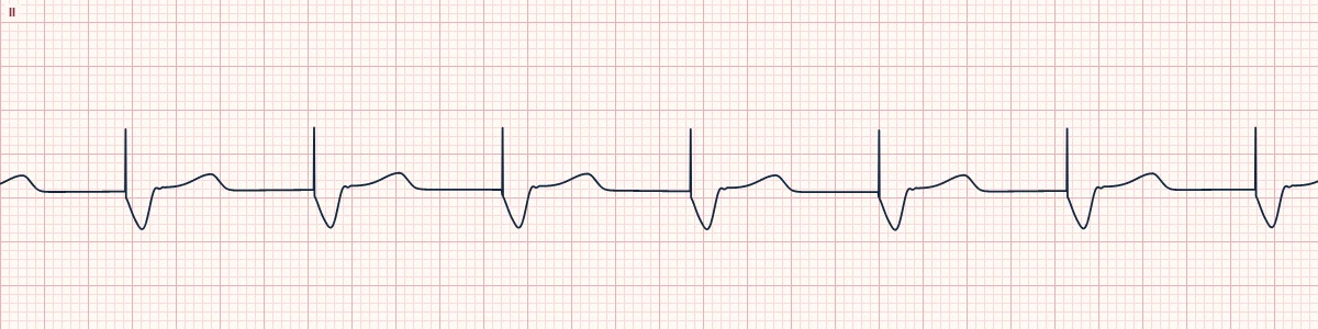
A pacemaker lead in the right ventricle starts each beat. Each pacing spike is followed by a wide QRS that resembles left bundle branch block.
What the heart is doing
The device stimulates the right ventricular apex. Activation spreads muscle to muscle from right to left and upward, so the left ventricle is activated late, mimicking LBBB.
Why it looks this way
This is a ventricular paced rhythm because a pacing spike immediately precedes each wide, LBBB-like QRS at a fixed regular rate.
Why it matters
Normal device function. Discordant ST–T changes are expected and hide infarction. Use the Sgarbossa criteria as modified by Dr. Stephen W. Smith: concordant ST elevation, V1–V3 ST depression of 1 mm or more, or discordant elevation at least 25% of the S-wave depth suggests occlusion.
What to look for
- A sharp vertical spike immediately followed by a wide QRS.
- The QRS is mostly negative in V1 and in the inferior leads, as expected from a right ventricular apical lead.
- Every spike must be followed by a QRS, and every QRS should produce a pulse.
Frequently asked questions
What are the ECG criteria for ventricular paced rhythm?
Rate: the programmed rate, perfectly regular. Pacing spike: one, immediately before each QRS. QRS: wide (≥ 120 ms), LBBB-like. Axis: leftward and superior (negative in II, III, aVF). ST–T: opposite to the main QRS deflection.
How is ventricular paced rhythm identified on an ECG?
This is a ventricular paced rhythm because a pacing spike immediately precedes each wide, LBBB-like QRS at a fixed regular rate.
What can ventricular paced rhythm be mistaken for?
Left bundle branch block: no spikes; a P wave precedes each QRS. Accelerated idioventricular rhythm: wide and regular but without spikes. Hyperkalemia: wide QRS with peaked T waves and no spikes.
What causes ventricular paced rhythm?
Common causes: complete or high-grade AV block; atrial fibrillation with a slow ventricular rate; symptomatic bradycardia; and temporary pacing during emergencies.