
Ventricular
Draft — pending owner review
Ventricular tachycardia, LBBB morphology
Ventricular tachycardia whose complexes look like left bundle branch block: mainly negative in V1. It starts in the right ventricle or septum.
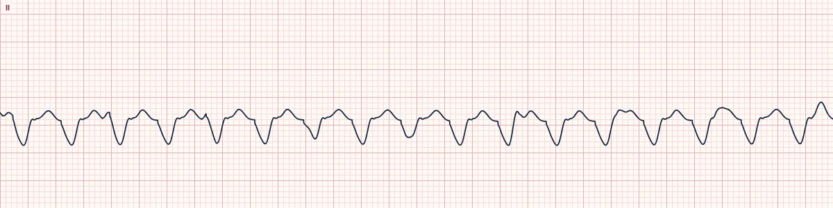
What the heart is doing
Activation begins in the right ventricle and spreads to the left through muscle rather than the bundles, so the main force points away from V1, as in left bundle branch block.
Why it looks this way
This is ventricular tachycardia with LBBB morphology because a fast, regular, wide rhythm is negative in V1 and the P waves are dissociated from it.
Why it matters
Treat as VT: assess for a pulse and perfusion first. LBBB-morphology VT with a superior axis in a young person should raise arrhythmogenic right ventricular cardiomyopathy and needs imaging.
What to look for
- Check V1: a broad negative complex means the impulse starts on the right side.
- Note the axis: superior with LBBB morphology suggests the right ventricular body or apex.
- Search for AV dissociation, capture or fusion beats.
Frequently asked questions
What are the ECG criteria for ventricular tachycardia, LBBB morphology?
Rate: usually 120–250/min. Rhythm: regular. QRS: wide (≥ 120 ms), dominant S wave in V1. Axis: superior axis points to the right ventricular body or apex. P waves: dissociated, often hidden.
How is ventricular tachycardia, LBBB morphology identified on an ECG?
This is ventricular tachycardia with LBBB morphology because a fast, regular, wide rhythm is negative in V1 and the P waves are dissociated from it.
What can ventricular tachycardia, LBBB morphology be mistaken for?
SVT with left bundle branch block: a typical LBBB shape with a rapid initial downstroke in V1. Monomorphic ventricular tachycardia (RBBB-like): dominant R in V1, left ventricular origin. Antidromic AVRT: fully pre-excited QRS.
What causes ventricular tachycardia, LBBB morphology?
Common causes: arrhythmogenic right ventricular cardiomyopathy; scar from septal or right ventricular infarction; sarcoidosis; repaired congenital heart disease; and right ventricular outflow tract focus (inferior axis, structurally normal heart).