
Ventricular
Multifocal PVCs
PVCs of two or more different shapes, meaning they arise from more than one site in the ventricles.
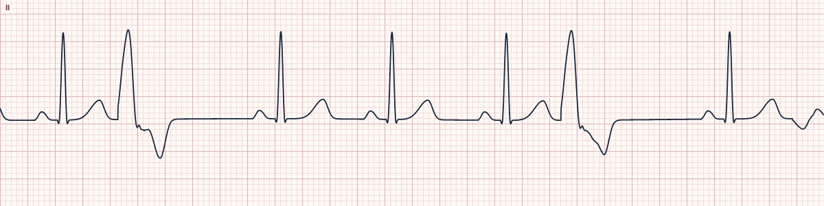
What the heart is doing
Several ventricular sites fire early at different times. Each site activates the ventricles along a different path, so each produces its own QRS shape and axis.
Why it looks this way
These are multifocal PVCs because the wide premature beats have at least two clearly different shapes.
Why it matters
Suggests widespread ventricular irritability and carries more risk than uniform PVCs. In an acutely ill patient, urgently look for ischemia, hypoxia and electrolyte loss.
What to look for
- Compare the PVCs in a single lead: some point up, some down, with different widths.
- Each shape tends to have its own coupling interval to the preceding beat.
- Check V1: upright PVCs come from the left ventricle, negative ones from the right.
Frequently asked questions
What are the ECG criteria for multifocal PVCs?
Rate: that of the underlying rhythm. Rhythm: irregular because of early beats. P waves: none before the early beats. QRS: wide PVCs of at least 2 distinct shapes. Coupling: interval to the PVC varies with its shape.
How is multifocal PVCs identified on an ECG?
These are multifocal PVCs because the wide premature beats have at least two clearly different shapes.
What can multifocal PVCs be mistaken for?
Unifocal PVCs with fusion beats: the odd shapes are blends with a sinus beat. Aberrantly conducted PACs: preceded by early P waves. Polymorphic VT: consecutive, not isolated, changing beats.
What causes multifocal PVCs?
Common causes: myocardial ischemia or infarction; hypokalemia, hypomagnesemia; hypoxia and acidosis; cardiomyopathy and heart failure; digoxin toxicity; and stimulant toxicity.