
Ventricular
Non-sustained ventricular tachycardia
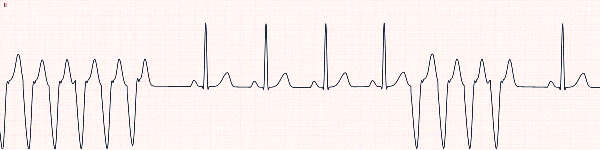
A short burst of ventricular tachycardia: three or more ventricular beats in a row faster than 100/min that stops by itself within 30 seconds.
What the heart is doing
A re-entry circuit around scar, or a rapidly firing ventricular focus, takes over for a few beats. It then extinguishes spontaneously and the sinus node resumes control.
Why it looks this way
This is non-sustained VT because three or more consecutive wide ventricular beats run faster than 100/min and stop spontaneously in under 30 seconds.
Why it matters
A warning sign, especially with structural heart disease, where it predicts sustained VT and sudden death. Correct electrolytes and ischemia, monitor continuously and arrange cardiac evaluation.
What to look for
- A sudden salvo of wide beats that interrupts sinus rhythm and then stops spontaneously.
- Count the beats: three or more consecutive ventricular beats.
- Note the patient's symptoms and pulse during the run.
Frequently asked questions
What are the ECG criteria for non-sustained ventricular tachycardia?
Rate: > 100/min during the run (here about 170). Duration: ≥ 3 beats, ending within 30 seconds. Rhythm: regular during the run. P waves: none related to the wide beats. QRS: wide (≥ 120 ms), uniform during the run.
How is non-sustained ventricular tachycardia identified on an ECG?
This is non-sustained VT because three or more consecutive wide ventricular beats run faster than 100/min and stop spontaneously in under 30 seconds.
What can non-sustained ventricular tachycardia be mistaken for?
SVT with aberrancy: starts with an early P wave and has a typical bundle branch shape. Artifact: sinus QRS complexes can be marched through the 'run'. Accelerated idioventricular rhythm: wide run at under about 110/min.
What causes non-sustained ventricular tachycardia?
Common causes: myocardial ischemia or prior infarction; cardiomyopathy and heart failure; hypokalemia, hypomagnesemia; QT-prolonging drugs; stimulants; and inherited arrhythmia syndromes.