
Arrest rhythms
Agonal rhythm
The last electrical activity of a dying heart: occasional, very slow, wide, misshapen complexes with no effective contraction.
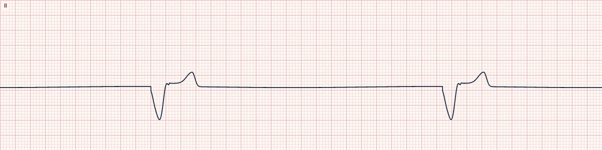
What the heart is doing
A failing ventricular focus fires occasionally and the impulse creeps slowly through energy-starved muscle. The electrical signal produces no useful mechanical contraction.
Why it looks this way
This is an agonal rhythm because the complexes are very wide and slurred, irregular, slower than 20/min, and have no P waves.
Why it matters
A pulseless arrest rhythm managed like asystole: continue CPR and treat reversible causes. Defibrillation is not indicated. It usually progresses to asystole.
What to look for
- Sparse, broad, sloppy complexes separated by long flat stretches.
- Complexes tend to get wider, lower and further apart over time.
- There is no pulse; do not wait for the monitor to go flat before acting.
Frequently asked questions
What are the ECG criteria for agonal rhythm?
Rate: < 20/min. Rhythm: irregular, slowing. P waves: absent. QRS: very wide, low, slurred complexes. Pulse: none.
How is agonal rhythm identified on an ECG?
This is an agonal rhythm because the complexes are very wide and slurred, irregular, slower than 20/min, and have no P waves.
What can agonal rhythm be mistaken for?
Idioventricular rhythm: regular at 20–40/min and may have a pulse. Asystole: no complexes at all. Severe hyperkalemia: wide sine-wave complexes, usually at a faster rate.
What causes agonal rhythm?
Common causes: prolonged cardiac arrest; severe hypoxia; profound acidosis; massive myocardial infarction; exsanguination; and end-stage illness.