
Arrest rhythms
Ventricular fibrillation, coarse
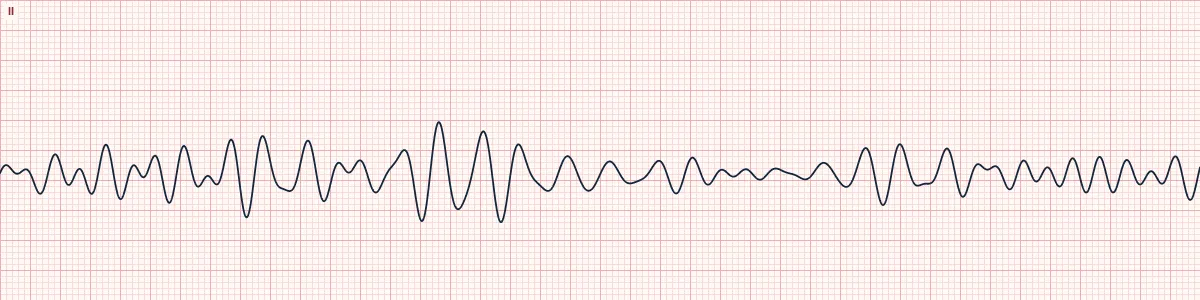
Chaotic electrical activity in the ventricles with no coordinated contraction and no pulse. Coarse VF has large waves and is the most shockable form.
What the heart is doing
Many wandering wavelets depolarize small patches of ventricle at random. With no organized wavefront the muscle only quivers, so there is no ejection and no cardiac output.
Why it looks this way
This is coarse ventricular fibrillation because the trace is chaotic with large irregular waves and no identifiable P waves, QRS complexes or T waves.
Why it matters
Cardiac arrest. Survival falls with every minute of delay: start high-quality CPR at once and defibrillate as early as possible, then continue the arrest algorithm and look for reversible causes.
What to look for
- A wandering, chaotic line with no repeating shape and no recognizable QRS or T wave.
- Wave height and width change constantly.
- Confirm the patient is unresponsive and pulseless; a loose lead can look similar.
Frequently asked questions
What are the ECG criteria for ventricular fibrillation, coarse?
Rate: not measurable. Rhythm: chaotic, completely irregular. P waves: none. QRS: none; no identifiable complexes. Waves: irregular undulations ≥ 3 mm tall.
How is ventricular fibrillation, coarse identified on an ECG?
This is coarse ventricular fibrillation because the trace is chaotic with large irregular waves and no identifiable P waves, QRS complexes or T waves.
What can ventricular fibrillation, coarse be mistaken for?
Artifact from movement or a loose electrode: the patient is awake with a pulse. Polymorphic VT or torsades: discrete wide complexes can still be made out. Fine VF: the same chaos at low amplitude.
What causes ventricular fibrillation, coarse?
Common causes: acute myocardial infarction or ischemia; degeneration of ventricular tachycardia; cardiomyopathy; severe electrolyte disturbance; electrocution, drowning, hypothermia; and inherited arrhythmia syndromes.