
Arrest rhythms
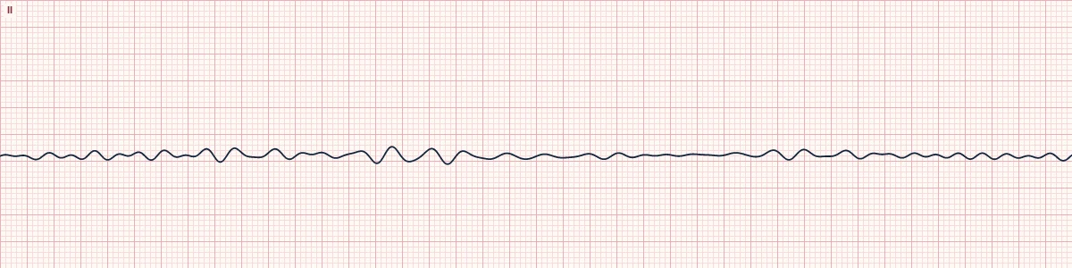
Ventricular fibrillation, fine
Ventricular fibrillation with low-amplitude waves. It usually means VF has been present longer and the heart is running out of energy.
What the heart is doing
The same disorganized wavelets as coarse VF, but the energy-depleted ventricle generates weaker signals. Without perfusion, the amplitude keeps falling until all activity stops.
Why it looks this way
This is fine ventricular fibrillation because the baseline is chaotic and low in amplitude with no P waves, QRS complexes or T waves.
Why it matters
Still a shockable arrest rhythm: CPR and defibrillation without delay. It converts less readily than coarse VF, and good chest compressions improve the odds of a successful shock.
What to look for
- A low, shivering baseline that never becomes truly flat.
- Check a second lead and increase the gain before calling it asystole.
- Make sure the electrodes and cables are attached.
Frequently asked questions
What are the ECG criteria for ventricular fibrillation, fine?
Rate: not measurable. Rhythm: chaotic, completely irregular. P waves: none. QRS: none. Waves: irregular undulations < 3 mm tall.
How is ventricular fibrillation, fine identified on an ECG?
This is fine ventricular fibrillation because the baseline is chaotic and low in amplitude with no P waves, QRS complexes or T waves.
What can ventricular fibrillation, fine be mistaken for?
Asystole: a nearly flat line in more than one lead at normal gain. Artifact or a disconnected lead: check the patient and the connections. Coarse VF: the same pattern with waves 3 mm or taller.
What causes ventricular fibrillation, fine?
Common causes: prolonged untreated coarse VF; acute myocardial infarction; severe hypoxia and acidosis; electrolyte disturbance; hypothermia; and drug toxicity.