
Arrest rhythms
Ventricular standstill (P-wave asystole)
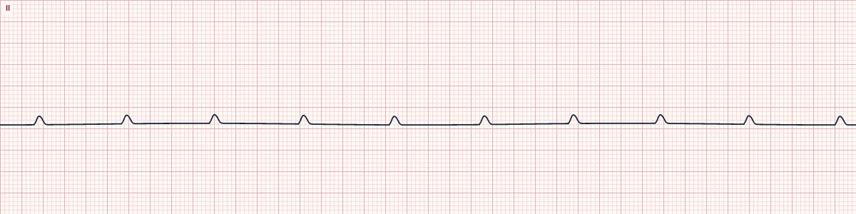
The atria keep beating under sinus control, but no impulse reaches the ventricles and no escape rhythm appears. There are P waves only, and no pulse.
What the heart is doing
The sinus node and atria work normally, but conduction is completely blocked below them and no junctional or ventricular focus takes over. The ventricles stay electrically silent.
Why it looks this way
This is ventricular standstill because regular P waves continue but there are no QRS complexes at all.
Why it matters
Cardiac arrest: start CPR immediately. Unlike true asystole, the ventricles may still respond to electrical pacing, so recognizing the P waves matters.
What to look for
- Small regular bumps on an otherwise flat line: P waves with nothing after them.
- No QRS complexes and no T waves anywhere.
- The patient is pulseless; treat the patient, not the reassuring-looking P waves.
Frequently asked questions
What are the ECG criteria for ventricular standstill (P-wave asystole)?
Rate: atrial 40–110/min; ventricular zero. Rhythm: P waves regular. P waves: present, normal shape. PR interval: none. QRS: absent.
How is ventricular standstill (P-wave asystole) identified on an ECG?
This is ventricular standstill because regular P waves continue but there are no QRS complexes at all.
What can ventricular standstill (P-wave asystole) be mistaken for?
Asystole: no P waves either. Third-degree AV block: slow QRS complexes are present among the P waves. Artifact: regular bumps from breathing or movement in a patient with a pulse.
What causes ventricular standstill (P-wave asystole)?
Common causes: complete AV block with failure of escape pacemakers; sudden progression of Mobitz II block; anterior myocardial infarction; severe hyperkalemia; and conduction system disease.