
AV blocks
Third-degree AV block, ventricular escape
No atrial impulse reaches the ventricles and the only backup is a slow ventricular pacemaker. The QRS is wide and the rate is 20–40/min.
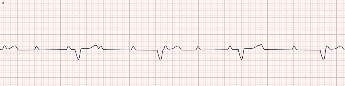
What the heart is doing
Conduction is interrupted below the His bundle. The sinus node still drives the atria, but the ventricles depend on a slow focus in the ventricular muscle that spreads cell to cell.
Why it looks this way
This is third-degree AV block with ventricular escape because regular P waves are unrelated to a slow, regular, wide QRS rhythm at 20–40/min.
Why it matters
An emergency. A ventricular escape is slow, unreliable and may stop without warning; atropine rarely helps. Support perfusion and move quickly to pacing; a permanent pacemaker is needed.
What to look for
- Slow, wide, regular QRS complexes with P waves scattered at a faster regular rate.
- No P wave has a consistent relationship to any QRS.
- Look for P waves deforming the T waves and ST segments.
Frequently asked questions
What are the ECG criteria for third-degree AV block, ventricular escape?
Rate: atrial 60–100/min; ventricular 20–40/min. Rhythm: P–P regular and R–R regular, but unrelated. P waves: normal; march through QRS and T waves. PR interval: varies randomly (no true PR). QRS: wide (≥ 120 ms).
How is third-degree AV block, ventricular escape identified on an ECG?
This is third-degree AV block with ventricular escape because regular P waves are unrelated to a slow, regular, wide QRS rhythm at 20–40/min.
What can third-degree AV block, ventricular escape be mistaken for?
Third-degree block with junctional escape: narrow QRS at 40–60/min. Sinus bradycardia with bundle branch block: one P wave before each QRS at a fixed PR. Idioventricular rhythm: no P waves at all.
What causes third-degree AV block, ventricular escape?
Common causes: anterior myocardial infarction; degenerative fibrosis of the conduction system; after cardiac surgery or valve procedures; hyperkalemia; drug toxicity; and infiltrative disease.