
AV blocks
2:1 AV block
Every second P wave is blocked, so there are two P waves for each QRS. Because no two conducted beats are consecutive, it cannot be labeled Mobitz I or II from the strip alone.
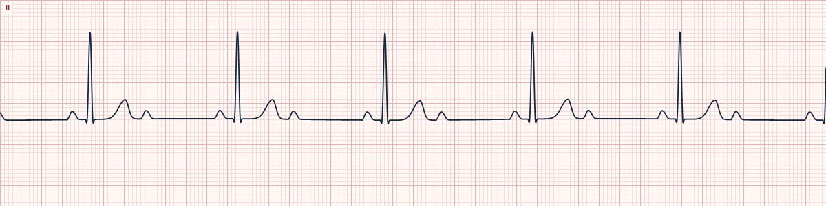
What the heart is doing
Alternate atrial impulses are blocked, either in the AV node or below it. A narrow QRS with a long PR favors the node; a wide QRS with a normal PR favors the His–Purkinje system.
Why it looks this way
This is 2:1 AV block because regular P waves outnumber QRS complexes two to one, with a constant PR on the conducted beats.
Why it matters
Treat with the caution due to Mobitz II until proven otherwise, particularly when the QRS is wide. Symptomatic patients are managed as for bradycardia with early preparation for pacing.
What to look for
- Look for an extra P wave midway between QRS complexes, often perched on the T wave.
- Check the P–P interval is regular, so the blocked P is not a premature beat.
- Watch a long strip for a stretch of 3:2 conduction that reveals the true type.
Frequently asked questions
What are the ECG criteria for 2:1 AV block?
Rate: ventricular exactly half the atrial rate. Rhythm: regular. P waves: 2 for each QRS; P–P interval regular. PR interval: constant on conducted beats. QRS: narrow suggests nodal block; wide suggests infranodal.
How is 2:1 AV block identified on an ECG?
This is 2:1 AV block because regular P waves outnumber QRS complexes two to one, with a constant PR on the conducted beats.
What can 2:1 AV block be mistaken for?
Sinus bradycardia: the blocked P wave is missed because it sits on the T wave. Non-conducted atrial bigeminy: the blocked P waves are early. Third-degree AV block: the PR varies and P waves have no relation to the QRS.
What causes 2:1 AV block?
Common causes: inferior or anterior myocardial infarction; beta-blockers, calcium-channel blockers, digoxin; degenerative conduction disease; myocarditis, Lyme disease; and high vagal tone.