
Sinus
Sinus bradycardia
A normal sinus rhythm that is simply slow, below 60/min. Often harmless, but it can reduce cardiac output when the rate is very low.
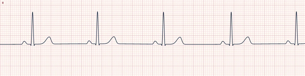
What the heart is doing
The sinus node discharges fewer than 60 times a minute, usually from vagal influence, drugs or ischemia. Each impulse still follows the normal pathway through the atria, AV node and ventricles.
Why it looks this way
This is sinus bradycardia because normal upright P waves precede every narrow QRS with a constant PR, but the rate is below 60/min.
Why it matters
Needs treatment only when it causes poor perfusion: hypotension, altered mental status, ischemic chest pain or shock. Correct hypoxia and other causes first; symptomatic patients may need atropine or pacing.
What to look for
- Everything looks like sinus rhythm except the rate: more than five large boxes between R waves.
- Confirm each P wave conducts with a fixed PR, so you are not missing an AV block.
- Search the T waves for hidden non-conducted P waves before settling on sinus bradycardia.
Frequently asked questions
What are the ECG criteria for sinus bradycardia?
Rate: < 60/min. Rhythm: regular. P waves: upright in II, one before every QRS. PR interval: 120–200 ms, constant. QRS: narrow (< 120 ms).
How is sinus bradycardia identified on an ECG?
This is sinus bradycardia because normal upright P waves precede every narrow QRS with a constant PR, but the rate is below 60/min.
What can sinus bradycardia be mistaken for?
2:1 AV block: a second P wave is hidden in or just after the T wave. Junctional escape rhythm: no upright P wave before the QRS. Non-conducted atrial bigeminy: an early P wave deforms every T wave.
What causes sinus bradycardia?
Common causes: sleep and athletic conditioning; high vagal tone (vomiting, straining); beta-blockers, calcium-channel blockers, digoxin; inferior myocardial infarction; hypoxia, hypothermia, hypothyroidism; and raised intracranial pressure.