
AV blocks
Second-degree AV block, Mobitz I (Wenckebach)
The AV node tires progressively: each PR is longer than the last until one P wave fails to conduct. The cycle then resets.
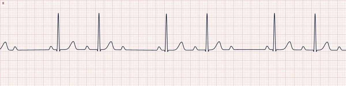
What the heart is doing
Each impulse reaches the AV node a little earlier in its recovery, so conduction slows further with every beat until one impulse is blocked. The rest lets the node recover and the cycle restarts.
Why it looks this way
This is Mobitz I because the PR interval lengthens progressively until a P wave is not followed by a QRS, then resets.
Why it matters
The block is in the AV node and is usually benign and transient; it rarely progresses to complete block. Treat only if the patient is symptomatic, and review rate-slowing drugs.
What to look for
- Find the dropped beat, then compare the PR before the pause (longest) with the PR after it (shortest).
- Beats cluster in groups of three, four or five separated by a pause.
- The R–R shortens before the dropped beat, and the pause is less than two cycles.
Frequently asked questions
What are the ECG criteria for second-degree AV block, Mobitz I (Wenckebach)?
Rate: atrial regular; ventricular slower. Rhythm: regularly irregular, grouped beating. P waves: regular; more P waves than QRS complexes. PR interval: lengthens beat by beat, then a P is not conducted. QRS: usually narrow.
How is second-degree AV block, Mobitz I (Wenckebach) identified on an ECG?
This is Mobitz I because the PR interval lengthens progressively until a P wave is not followed by a QRS, then resets.
What can second-degree AV block, Mobitz I (Wenckebach) be mistaken for?
Mobitz II: the PR is constant before and after the dropped beat. Non-conducted PACs: the blocked P wave is early and a different shape. Sinus arrhythmia: every P wave conducts.
What causes second-degree AV block, Mobitz I (Wenckebach)?
Common causes: high vagal tone (sleep, athletes); inferior myocardial infarction; beta-blockers, calcium-channel blockers, digoxin; myocarditis, Lyme disease; and after cardiac surgery.