
Electrolytes
Hypercalcemia
High blood calcium shortens the plateau of the action potential, so the ST segment almost disappears and the QT interval is short.
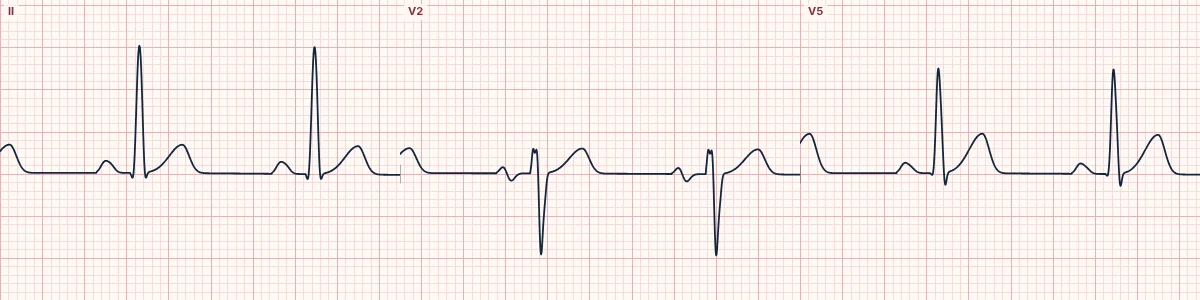
What the heart is doing
A high calcium level shortens phase 2 (the plateau) of the ventricular action potential. Because the plateau corresponds to the ST segment, the ST shortens and the QT with it.
Why it looks this way
This is hypercalcemia because the QT is short due to a nearly absent ST segment, while the T wave itself looks normal.
Why it matters
The ECG change is a clue to the diagnosis more than a danger in itself. Severe hypercalcemia causes dehydration, confusion and occasionally bradycardia or heart block; treat with fluids and the cause.
What to look for
- The T wave takes off almost straight from the end of the QRS.
- Measure the QT and correct it for rate: it is shorter than expected.
- T-wave shape and height are essentially normal.
Frequently asked questions
What are the ECG criteria for hypercalcemia?
QT: short (QTc < 360 ms). ST segment: very short or absent. T waves: begin right after the QRS; normal shape. Severe cases: PR prolongation, QRS widening, J waves.
How is hypercalcemia identified on an ECG?
This is hypercalcemia because the QT is short due to a nearly absent ST segment, while the T wave itself looks normal.
What can hypercalcemia be mistaken for?
Congenital short QT: tall, peaked T waves and a family history. Digoxin effect: short QT with a scooped ST segment. Hyperkalemia: peaked narrow T waves.
What causes hypercalcemia?
Common causes: hyperparathyroidism; cancer (bone metastases or hormone-secreting tumors); excess vitamin D or calcium intake; thiazide diuretics; and sarcoidosis and other granulomatous disease.