
Electrolytes
Hypokalemia
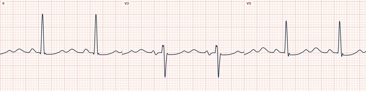
Low blood potassium delays repolarization: T waves flatten, the ST segment sags and a U wave appears after the T wave.
What the heart is doing
With less potassium outside the cells, the outward potassium current is weaker and repolarization is drawn out. The late part of repolarization shows up as a U wave.
Why it looks this way
This is hypokalemia because the T waves are flattened, the ST segment is mildly depressed and prominent U waves follow the T waves.
Why it matters
Makes the ventricles irritable: PVCs, ventricular tachycardia and torsades de pointes, especially in patients on digoxin or with heart disease. Replace potassium, and magnesium with it.
What to look for
- An extra bump after the T wave in V2–V3: the U wave.
- T waves become smaller as U waves become larger.
- A seemingly very long QT with a double-humped T wave is usually T–U fusion.
Frequently asked questions
What are the ECG criteria for hypokalemia?
T waves: flattened or inverted. U waves: prominent, best in V2–V3; may exceed the T wave. ST segment: mild depression. QT: appears long because T and U merge (QU interval).
How is hypokalemia identified on an ECG?
This is hypokalemia because the T waves are flattened, the ST segment is mildly depressed and prominent U waves follow the T waves.
What can hypokalemia be mistaken for?
Long QT syndrome: a single broad T wave with no separate U wave. Ischemia: ST depression without U waves, with symptoms. Digoxin effect: scooped ST with a short QT.
What causes hypokalemia?
Common causes: diuretics; vomiting and diarrhea; insulin or beta-agonist treatment; hypomagnesemia; and excess aldosterone or steroids.