
Atrial
Atrial fibrillation
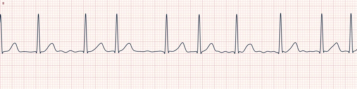
Chaotic electrical activity makes the atria quiver instead of contract. There are no P waves and the ventricles respond at random, here at a controlled rate.
What the heart is doing
Hundreds of disorganized wavelets circulate through the atria. The AV node is bombarded irregularly and passes only some impulses, each along the normal pathway, so the QRS is narrow but randomly timed.
Why it looks this way
This is atrial fibrillation because the rhythm is irregularly irregular with no P waves, only a fibrillatory baseline, and narrow QRS complexes.
Why it matters
Loss of atrial contraction reduces filling and lets clot form in the left atrium, raising stroke risk. Care centers on rate or rhythm control and anticoagulation based on stroke risk.
What to look for
- No two R–R intervals match and there is no repeating pattern.
- Search II and V1 for P waves; you will find only a wavy, shifting baseline.
- Count the rate over at least 6 seconds, because single R–R intervals mislead.
Frequently asked questions
What are the ECG criteria for atrial fibrillation?
Rate: ventricular 60–110/min (controlled). Rhythm: irregularly irregular. P waves: none; fine fibrillatory waves on the baseline. PR interval: not measurable. QRS: narrow (< 120 ms).
How is atrial fibrillation identified on an ECG?
This is atrial fibrillation because the rhythm is irregularly irregular with no P waves, only a fibrillatory baseline, and narrow QRS complexes.
What can atrial fibrillation be mistaken for?
Multifocal atrial tachycardia or wandering pacemaker: distinct P waves before each QRS. Atrial flutter with variable block: regular identical sawtooth waves. Sinus rhythm with frequent PACs: a dominant P wave can be found.
What causes atrial fibrillation?
Common causes: hypertension and aging; heart failure, coronary disease; mitral and other valve disease; hyperthyroidism; alcohol, sleep apnea; and acute illness, surgery, pulmonary embolism.