
Atrial
Ectopic atrial rhythm
A steady rhythm at a normal rate driven by an atrial focus other than the sinus node, recognized by an abnormal P-wave shape or axis.
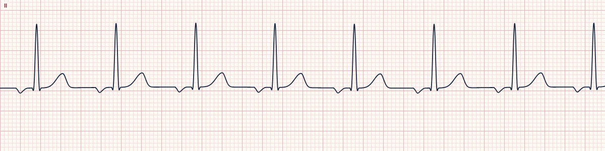
What the heart is doing
A focus low in the atrium fires slightly faster than the sinus node and takes over. The atria depolarize from bottom to top, inverting the P wave in the inferior leads; AV conduction is normal.
Why it looks this way
This is an ectopic atrial rhythm because inverted P waves with a PR of at least 120 ms precede every narrow QRS at a rate under 100/min.
Why it matters
Usually an incidental, harmless finding that needs no treatment. It matters mainly because it can be confused with lead misplacement or a junctional rhythm.
What to look for
- Check lead II first: a negative P wave before every QRS means the atria are being activated from below.
- Measure the PR. At 120 ms or more the focus is atrial; shorter than that suggests a junctional origin.
- Rule out misplaced limb electrodes before accepting an abnormal P axis.
Frequently asked questions
What are the ECG criteria for ectopic atrial rhythm?
Rate: < 100/min. Rhythm: regular. P waves: non-sinus: here inverted in II, III and aVF. PR interval: ≥ 120 ms, constant. QRS: narrow (< 120 ms).
How is ectopic atrial rhythm identified on an ECG?
This is an ectopic atrial rhythm because inverted P waves with a PR of at least 120 ms precede every narrow QRS at a rate under 100/min.
What can ectopic atrial rhythm be mistaken for?
Accelerated junctional rhythm: inverted P waves with PR < 120 ms, or none. Arm lead reversal: P, QRS and T are all inverted in lead I, not II. Normal sinus rhythm: P waves upright in II.
What causes ectopic atrial rhythm?
Common causes: normal variant, especially in the young and during sleep; high vagal tone slowing the sinus node; sinus node dysfunction; atrial enlargement or scarring; and digoxin effect.