
Atrial
Focal atrial tachycardia
A rapid regular rhythm fired by a single atrial focus outside the sinus node. Each abnormal P wave conducts to a narrow QRS.
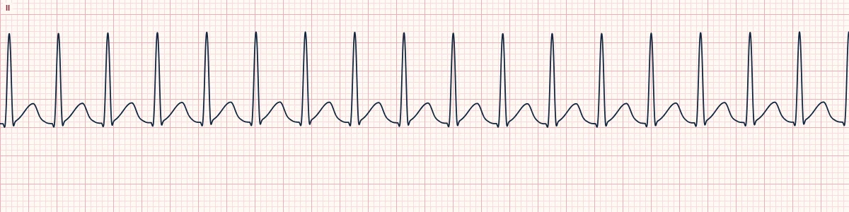
What the heart is doing
A single atrial focus fires rapidly, or a small circuit spins within the atrium. Impulses spread across the atria from that site and conduct through the AV node, usually 1:1.
Why it looks this way
This is focal atrial tachycardia because a regular narrow tachycardia has an abnormal P wave before every QRS, with a flat baseline between P waves.
Why it matters
Unstable patients need immediate synchronized cardioversion. In stable patients, vagal maneuvers or adenosine may only slow AV conduction and reveal the P waves; rate control and treating the cause follow.
What to look for
- Find a P wave before each QRS whose shape or axis differs from the patient's sinus P wave.
- A flat baseline between P waves separates it from flutter, which has no flat segment.
- It often warms up over a few beats at onset and may continue despite AV block.
Frequently asked questions
What are the ECG criteria for focal atrial tachycardia?
Rate: 100–250/min, regular. P waves: one abnormal shape before every QRS. Baseline: flat (isoelectric) between P waves. PR interval: ≥ 120 ms; the RP is longer than the PR. QRS: narrow (< 120 ms).
How is focal atrial tachycardia identified on an ECG?
This is focal atrial tachycardia because a regular narrow tachycardia has an abnormal P wave before every QRS, with a flat baseline between P waves.
What can focal atrial tachycardia be mistaken for?
Sinus tachycardia: normal upright P waves in II and gradual rate changes. Atrial flutter 2:1: sawtooth waves at about 300/min with no flat baseline. AVNRT: P wave hidden in or just after the QRS.
What causes focal atrial tachycardia?
Common causes: atrial scar after surgery or ablation; structural heart and lung disease; digoxin toxicity (often with AV block); catecholamine excess and stimulants; and idiopathic in healthy hearts.