
Atrial
Draft — pending owner review
Atrial tachycardia with 2:1 block
A fast ectopic atrial rhythm in which the AV node lets only every second P wave through. It is the classic rhythm of digoxin toxicity.
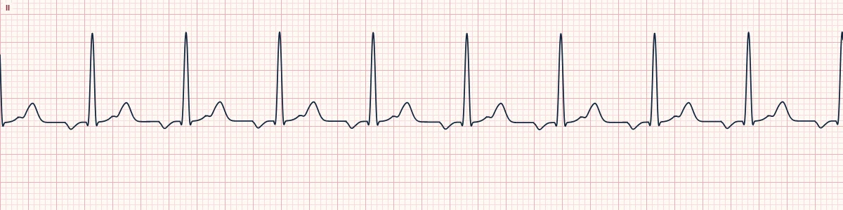
What the heart is doing
An ectopic atrial focus fires rapidly through enhanced automaticity or triggered activity. The AV node, slowed further by vagal or drug effects, blocks alternate impulses, so the ventricles beat at half the atrial rate.
Why it looks this way
This is atrial tachycardia with 2:1 block because abnormal P waves at about 180/min sit on a flat baseline and only every second one is followed by a QRS.
Why it matters
In a patient taking digoxin this rhythm is a red flag for toxicity: faster atrial firing together with slower AV conduction. Check the potassium and drug level and get expert advice; slowing the AV node further is rarely the answer.
What to look for
- Find the second P wave: it hides in the T wave or sits midway between QRS complexes.
- Check for a flat baseline between P waves; flutter waves never flatten.
- Ask about digoxin and check the potassium whenever this rhythm appears.
Frequently asked questions
What are the ECG criteria for atrial tachycardia with 2:1 block?
Atrial rate: 150–250/min, regular. P waves: abnormal in shape, with a flat baseline between them. Conduction: 2:1 AV block, sometimes variable. Ventricular rate: half the atrial rate. QRS: narrow unless there is bundle branch block.
How is atrial tachycardia with 2:1 block identified on an ECG?
This is atrial tachycardia with 2:1 block because abnormal P waves at about 180/min sit on a flat baseline and only every second one is followed by a QRS.
What can atrial tachycardia with 2:1 block be mistaken for?
Atrial flutter 2:1: sawtooth waves with no flat baseline and an atrial rate near 300. Sinus tachycardia: one upright P wave before every QRS. 2:1 AV block: sinus P waves at a normal atrial rate.
What causes atrial tachycardia with 2:1 block?
Common causes: digoxin toxicity; low potassium, which worsens digoxin toxicity; structural heart disease; chronic lung disease; and atrial scarring or prior atrial surgery.