
Atrial
Multifocal atrial tachycardia
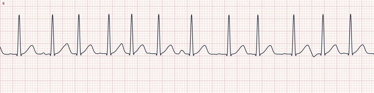
A fast, irregular rhythm with P waves of at least three shapes, driven by several irritable atrial foci. Classically seen in severe lung disease.
What the heart is doing
Multiple atrial sites fire spontaneously and compete, usually because of hypoxia, atrial stretch or electrolyte loss. Each impulse takes a different route to the AV node, giving chaotic timing.
Why it looks this way
This is multifocal atrial tachycardia because the rate exceeds 100/min, the rhythm is irregular, and there are at least three P-wave shapes with varying PR.
Why it matters
A marker of a sick patient rather than a primary rhythm problem. Treat the cause: oxygenation, ventilation and electrolytes. Cardioversion does not work because there is no single circuit to interrupt.
What to look for
- An irregular tachycardia that still has a clear P wave before each QRS.
- Count P-wave shapes in a single lead; three or more confirm it.
- Look for a flat baseline between P waves, unlike the chaotic baseline of atrial fibrillation.
Frequently asked questions
What are the ECG criteria for multifocal atrial tachycardia?
Rate: > 100/min. Rhythm: irregularly irregular. P waves: at least 3 different shapes in one lead. PR interval: varies; some P waves may not conduct. QRS: narrow (< 120 ms).
How is multifocal atrial tachycardia identified on an ECG?
This is multifocal atrial tachycardia because the rate exceeds 100/min, the rhythm is irregular, and there are at least three P-wave shapes with varying PR.
What can multifocal atrial tachycardia be mistaken for?
Atrial fibrillation: no discrete P waves; fibrillatory baseline. Atrial flutter with variable block: identical sawtooth waves at about 300/min. Sinus tachycardia with frequent PACs: one dominant P-wave shape.
What causes multifocal atrial tachycardia?
Common causes: COPD exacerbation and respiratory failure; hypoxia; pulmonary hypertension; hypokalemia and hypomagnesemia; sepsis and critical illness; and theophylline or beta-agonist excess.