
Overviews
Draft — pending owner review
Can't-miss ECG patterns
The tracings that change what happens next
A short list of patterns that need action whatever the patient looks like: occlusion MI and its equivalents, dangerous rhythms, pre-excited atrial fibrillation, toxic and electrolyte patterns, and inherited causes of sudden death.
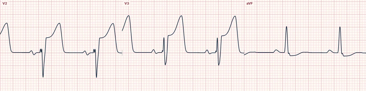
Occlusion and its disguises
ST elevation in contiguous leads, and also the equivalents that miss STEMI criteria: hyperacute T waves, de Winter T waves, posterior MI (ST depression deepest in V1–V3), the Aslanger and South African flag patterns, and occlusion through LBBB or pacing judged by the modified Sgarbossa criteria. Wellens T waves mean a critical LAD lesion that has reopened for now.
Rhythms that cannot wait
Ventricular tachycardia and ventricular flutter, polymorphic VT and torsades de pointes, pre-excited atrial fibrillation, Mobitz II, high-grade and complete AV block, and bifascicular or alternating bundle branch block in a patient who has fainted.
Poisons and electrolytes
Peaked T waves, a widening QRS or a sine wave in hyperkalemia; a wide QRS with a terminal R wave in aVR after a sodium-channel blocker overdose; a QTc above 500 ms; atrial tachycardia with block or bidirectional VT in digoxin toxicity.
Inherited and structural
Brugada type 1, the WPW pattern, long and short QT, hypertrophic cardiomyopathy, arrhythmogenic right ventricular cardiomyopathy (epsilon waves, T inversion in V1–V3) and the right heart strain of pulmonary embolism. In a patient with syncope, each of these changes the plan.
How to read it
- Read the whole ECG systematically first, then run this list deliberately before calling it normal.
- Compare with an old ECG and repeat the tracing when symptoms change.
- Match the ECG to the patient: syncope, chest pain or an overdose raise the stakes of every finding.
Pitfalls
- A computer reading of normal does not exclude any of these patterns.
- One ECG is a snapshot: occlusion, hyperkalemia and ischemia evolve over minutes.
Frequently asked questions
Can't-miss ECG patterns: what is the key idea?
A short list of patterns that need action whatever the patient looks like: occlusion MI and its equivalents, dangerous rhythms, pre-excited atrial fibrillation, toxic and electrolyte patterns, and inherited causes of sudden death.
Can't-miss ECG patterns: what are the key numbers?
Act now: Occlusion MI or equivalent, VT, pre-excited AF, high-grade AV block; QTc: > 500 ms: high risk of torsades; QRS after an overdose: > 100 ms: sodium-channel blockade; Syncope plus: Brugada, WPW, long QT, HCM or bifascicular block needs a full workup.
Can't-miss ECG patterns: how do you apply it to an ECG?
Read the whole ECG systematically first, then run this list deliberately before calling it normal. Compare with an old ECG and repeat the tracing when symptoms change. Match the ECG to the patient: syncope, chest pain or an overdose raise the stakes of every finding.
Can't-miss ECG patterns: what are the common pitfalls?
A computer reading of normal does not exclude any of these patterns. One ECG is a snapshot: occlusion, hyperkalemia and ischemia evolve over minutes.