
Overviews
Draft — pending owner review
Occlusion MI (OMI) and the limits of STEMI criteria
Looking for the occluded artery, not a millimeter threshold
Acute coronary occlusion is what needs emergency reperfusion, and many occlusions never meet millimeter STEMI criteria. The occlusion MI approach reads each ECG for signs of an occluded artery rather than for a threshold alone.
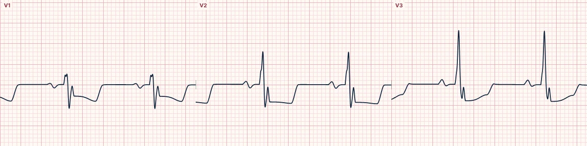
Why thresholds miss occlusions
STEMI criteria measure ST elevation at the J point against fixed cutoffs. A small QRS carries a small ST shift, the posterior and high lateral walls are poorly seen by the standard leads, and the earliest change is often in the T wave. Each of these can hide an occluded artery. Studies find an occluded culprit artery in roughly one in four patients labeled as non-ST-elevation MI.
Signs of occlusion below the thresholds
Hyperacute T waves; ST elevation that is small in millimeters but large for its QRS; reciprocal ST depression, especially in aVL with inferior changes; ST depression deepest in V1–V4 from posterior MI; the de Winter, Aslanger and South African flag patterns; terminal QRS distortion; and positive Sgarbossa–Smith criteria in LBBB or pacing.
Signs that argue against occlusion
ST elevation that is concave, in proportion to a tall QRS and unchanged on repeat tracings, as in early repolarization; ST elevation over QS waves with small T waves, as in an old aneurysm; and the proportionate secondary changes of LVH or bundle branch block.
Using it in practice
Local STEMI criteria still drive cath lab activation in most systems. Use the occlusion approach to escalate: repeat the ECG every 15–30 minutes, add posterior and right-sided leads, compare with old tracings and call early when the ECG and the story point to an occluded artery.
How to read it
- Read the ST segment and T wave as a shape, in proportion to the QRS, not only in millimeters.
- Check aVL on every inferior ECG and think posterior MI with every anterior ST depression.
- Repeat the ECG while pain continues: an occlusion evolves.
Pitfalls
- No STEMI does not mean no occlusion.
- Occlusion MI language is not yet universal; describe the findings in terms your receiving team acts on.
Frequently asked questions
Occlusion MI (OMI) and the limits of STEMI criteria: what is the key idea?
Acute coronary occlusion is what needs emergency reperfusion, and many occlusions never meet millimeter STEMI criteria. The occlusion MI approach reads each ECG for signs of an occluded artery rather than for a threshold alone.
Occlusion MI (OMI) and the limits of STEMI criteria: what are the key numbers?
STEMI threshold V2–V3: ≥ 2 mm men ≥ 40, ≥ 2.5 mm men < 40, ≥ 1.5 mm women; STEMI threshold other leads: ≥ 1 mm in two contiguous leads; Posterior leads V7–V9: ≥ 0.5 mm; Occluded artery in NSTEMI: Roughly 1 in 4 at angiography.
Occlusion MI (OMI) and the limits of STEMI criteria: how do you apply it to an ECG?
Read the ST segment and T wave as a shape, in proportion to the QRS, not only in millimeters. Check aVL on every inferior ECG and think posterior MI with every anterior ST depression. Repeat the ECG while pain continues: an occlusion evolves.
Occlusion MI (OMI) and the limits of STEMI criteria: what are the common pitfalls?
No STEMI does not mean no occlusion. Occlusion MI language is not yet universal; describe the findings in terms your receiving team acts on.