
Coronary occlusion (STEMI)
Posterior branch occlusion
Isolated posterior MI
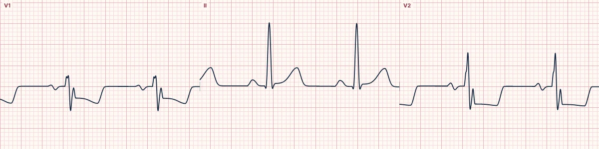
Blockage of a posterior branch, usually of the left circumflex artery (LCx), infarcting the posterior wall. No standard lead faces it, so it shows only as mirror-image changes in V1–V3.
What the heart is doing
Posterior wall injury produces an ST vector pointing straight backward, away from the chest leads. V1–V3 record it upside down: ST depression for elevation and tall R waves for Q waves.
Why it looks this way
This is a posterior occlusion because ST depression with tall R waves in V1–V3 mirrors ST elevation recorded in the posterior leads V7–V9.
Why it matters
Frequently mislabeled as non-ST-elevation ischemia, delaying reperfusion. ST depression maximal in V1–V3 with symptoms should be treated as a STEMI equivalent and confirmed with posterior leads.
What to look for
- Horizontal ST depression greatest in V1–V3 should always prompt posterior leads.
- A tall, broad R wave in V2 (R larger than S) is a posterior Q wave seen from the front.
- Place V7–V9 on the back at the level of V6; small elevations count.
Frequently asked questions
What are the ECG criteria for posterior branch occlusion?
Artery: distal circumflex (posterior branch). ST elevation: V7, V8, V9. Reciprocal ST depression: V1, V2, V3. V1–V3: horizontal ST depression and a tall, broad R; T upright once established. Threshold: ≥ 0.5 mm of elevation in V7–V9 is significant.
How is posterior branch occlusion identified on an ECG?
This is a posterior occlusion because ST depression with tall R waves in V1–V3 mirrors ST elevation recorded in the posterior leads V7–V9.
What can posterior branch occlusion be mistaken for?
Anterior subendocardial ischemia: depression usually maximal in V4–V6. RBBB: rsR′ in V1 with a wide QRS. Right ventricular hypertrophy: tall R in V1 with right axis deviation.
What causes posterior branch occlusion?
Common causes: plaque rupture with thrombosis; coronary artery spasm or stimulant use; spontaneous coronary dissection; and coronary embolism.