
Coronary occlusion (STEMI)
Mid LAD occlusion
Anterior STEMI
Blockage of the left anterior descending artery (LAD) beyond its first major branches. It infarcts the anterior wall and apex of the left ventricle.
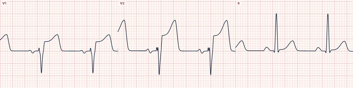
What the heart is doing
Transmural ischemia of the anterior wall and apex creates an injury current pointing straight toward the front of the chest, raising the ST segment in the precordial leads over it.
Why it looks this way
This is a mid LAD occlusion because ST elevation is confined to the precordial leads V1–V5 without high lateral involvement or reciprocal inferior depression.
Why it matters
A time-critical STEMI threatening a large part of the left ventricle. Immediate reperfusion is the priority; anticipate ventricular arrhythmias and pump failure.
What to look for
- ST elevation is greatest in V2–V4, directly over the anterior wall.
- I, aVL and the inferior leads are spared, placing the blockage beyond the first diagonal.
- Compare T-wave size with the QRS: a T wave towering over a small R is hyperacute.
Frequently asked questions
What are the ECG criteria for mid LAD occlusion?
Artery: mid LAD. ST elevation: V1, V2, V3, V4, V5. Reciprocal ST depression: none expected; inferior and high lateral leads stay near baseline. T waves: tall, broad hyperacute T waves, maximal V2–V4. QRS: Q waves and loss of R waves develop in V2–V4.
How is mid LAD occlusion identified on an ECG?
This is a mid LAD occlusion because ST elevation is confined to the precordial leads V1–V5 without high lateral involvement or reciprocal inferior depression.
What can mid LAD occlusion be mistaken for?
Early repolarization: notched J point, concave ST, T wave not broad. Left ventricular hypertrophy or LBBB: ST elevation in V1–V3 proportional to a deep S wave. Brugada pattern: coved elevation limited to V1–V2.
What causes mid LAD occlusion?
Common causes: plaque rupture with thrombosis; coronary artery spasm or stimulant use; spontaneous coronary dissection; coronary embolism; and stent thrombosis.