
Bundle branch block
Right bundle branch block
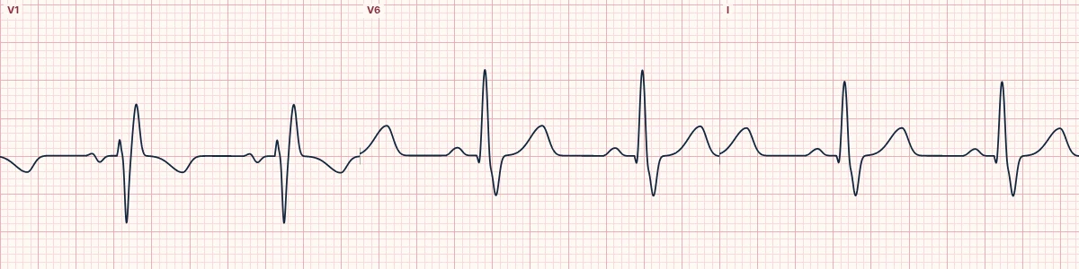
The right bundle branch does not conduct, so the right ventricle is activated late, through muscle, from the left side. The QRS widens with a late rightward, anterior force.
What the heart is doing
The left ventricle activates on time through the left bundle. The impulse then crosses the septum and spreads slowly through right ventricular muscle, adding a late force toward V1.
Why it looks this way
This is RBBB because the QRS is at least 120 ms with an rsR′ in V1 and a wide slurred S wave in I and V6.
Why it matters
Often benign when isolated. A new RBBB with chest pain or breathlessness suggests proximal LAD occlusion or pulmonary embolism. RBBB does not hide ST-elevation infarction.
What to look for
- Look at V1: the QRS ends with a tall, late R′ wave.
- Look at I and V6: the QRS ends with a broad, shallow S wave.
- The first part of the QRS is normal, so Q waves and ST elevation can still be read.
Frequently asked questions
What are the ECG criteria for right bundle branch block?
QRS: ≥ 120 ms. V1: rsR′ ('M' shape) or a broad notched R. V6 and I: wide, slurred S wave (> 40 ms). ST–T: T inversion in V1–V3 is expected. Axis: usually normal.
How is right bundle branch block identified on an ECG?
This is RBBB because the QRS is at least 120 ms with an rsR′ in V1 and a wide slurred S wave in I and V6.
What can right bundle branch block be mistaken for?
Brugada pattern: coved ST elevation in V1–V2 without a wide S in V6. Posterior infarction or RVH: tall R in V1 with a narrow QRS. WPW type A: short PR and delta wave.
What causes right bundle branch block?
Common causes: normal variant; right ventricular strain, pulmonary embolism; chronic lung disease; ischemic heart disease, anterior infarction; congenital heart disease (atrial septal defect); and degenerative conduction disease.