
Artifact
Draft — pending owner review
Artifact mimicking VT or VF
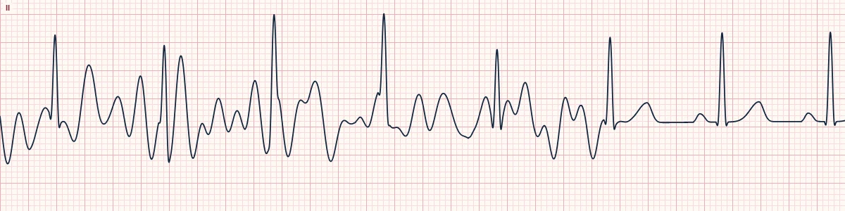
Electrical noise from patient movement, tremor or a loose electrode that looks like VT or VF on the monitor, while the heart is in an organized rhythm underneath.
What the heart is doing
The noise is not cardiac. A moving electrode changes its own potential, and every lead that uses that electrode records the change. The heart's activity continues underneath, so the real QRS complexes keep their timing.
Why it looks this way
This is artifact mimicking VT or VF because narrow QRS complexes keep marching through the noise at a steady rate, and the lead that does not use the moving electrode is clean.
Why it matters
Misread artifact leads to unneeded shocks, drugs and alarms. Always confirm a monitor rhythm against the patient and a pulse before treating; true VF needs immediate defibrillation and must not be dismissed as artifact.
What to look for
- Look at the patient first: VF does not talk or brush its teeth.
- Find the real QRS complexes and march them out with calipers through the noise.
- Check other leads: noise from one electrode spares the lead that does not use it.
- Compare with the pulse oximeter or arterial waveform, which keep a regular pulse.
Frequently asked questions
What are the ECG criteria for artifact mimicking VT or VF?
Patient: awake, talking or moving, with a pulse. Underlying QRS: narrow complexes keep marching through at a steady rate. Noise: irregular, often fastest and largest in some leads only. Clean lead: A lead that does not use the moving electrode looks normal. Rate: the rate of the hidden QRS matches the pulse.
How is artifact mimicking VT or VF identified on an ECG?
This is artifact mimicking VT or VF because narrow QRS complexes keep marching through the noise at a steady rate, and the lead that does not use the moving electrode is clean.
What can artifact mimicking VT or VF be mistaken for?
Ventricular fibrillation, coarse: no organized QRS anywhere and no pulse. Polymorphic ventricular tachycardia: wide complexes in every lead, no hidden narrow QRS. Torsades de pointes: twisting wide complexes, usually after a long QT.
What causes artifact mimicking VT or VF?
Common causes: tremor (shivering, Parkinson disease, seizures); patient movement, brushing teeth, chest physiotherapy; loose or dry electrodes; transport vibration; and electrical interference.