
Ischemia & prior infarction
Draft — pending owner review
Hyperacute T waves
The earliest ECG sign of a coronary occlusion: broad, bulky T waves that are tall for the size of their QRS, often before obvious ST elevation appears.
What the heart is doing
In the first minutes of occlusion, ischemic cells shorten their action potentials and leak potassium. The repolarization gradient over the injured wall steepens and the T wave grows before an injury current lifts the ST segment.
Why it looks this way
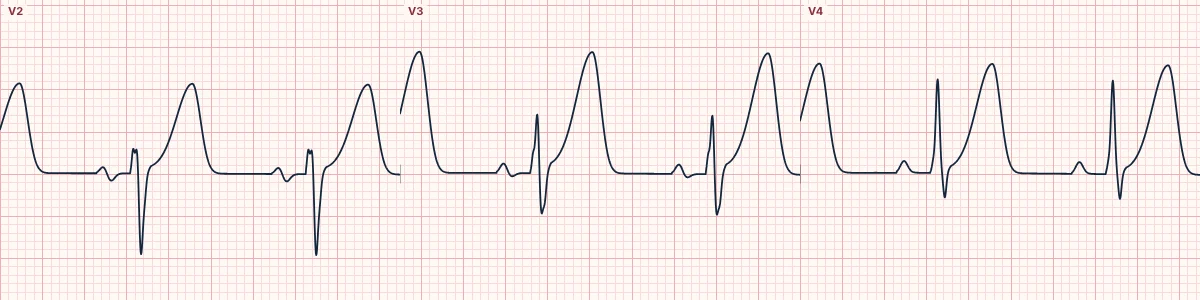
These are hyperacute T waves because V2–V4 show broad, bulky T waves out of proportion to the QRS with only slight ST elevation.
Why it matters
Hyperacute T waves can be the only sign of an occluded artery for the first minutes to hours. In the right setting treat them as an occlusion MI: repeat the ECG, involve a cardiologist early and do not wait for STEMI criteria.
What to look for
- Compare each T wave with its QRS: a bulky T over a modest QRS is suspicious.
- Look for a straight ST segment running up into a fat T wave.
- Repeat the ECG every 15–30 minutes while symptoms continue.
Frequently asked questions
What are the ECG criteria for hyperacute T waves?
T waves: broad-based, bulky and tall relative to the QRS. Leads: contiguous leads over one territory (here V2–V4). ST segment: straightened, or only slightly elevated. Evolution: often progress to ST elevation within minutes to hours. Comparison: new compared with a previous ECG.
How is hyperacute T waves identified on an ECG?
These are hyperacute T waves because V2–V4 show broad, bulky T waves out of proportion to the QRS with only slight ST elevation.
What can hyperacute T waves be mistaken for?
Hyperkalemia, mild: narrow, pointed, symmetric T waves in many leads. Benign early repolarization: T waves in proportion to a tall QRS, with a notched J point. De Winter T waves: upsloping ST depression leading into the tall T.
What causes hyperacute T waves?
Common causes: acute coronary occlusion, most often the LAD; re-occlusion after spontaneous reperfusion; and coronary artery spasm.