
Ischemia & prior infarction
Prior inferior infarction
The lasting scar of an earlier inferior wall infarct. Dead muscle produces no electrical force, leaving deep Q waves in the inferior leads.
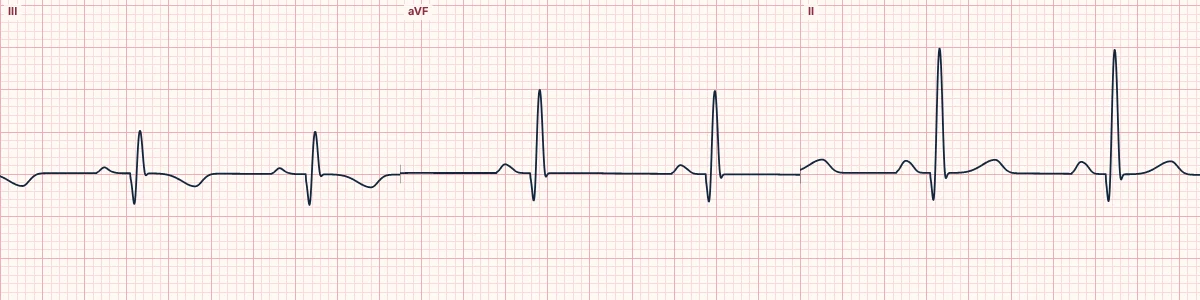
What the heart is doing
Scarred inferior wall generates no depolarization force. The inferior leads then record the opposite wall's forces moving away from them at the start of the QRS, inscribing a Q wave.
Why it looks this way
This is a prior inferior infarction because pathological Q waves are present in II, III and aVF with the ST segment back at baseline.
Why it matters
Evidence of established coronary disease and lost muscle. Not an emergency in its own right, but it raises the likelihood that new symptoms are cardiac; compare with previous ECGs.
What to look for
- Wide, deep Q waves in at least two of the three inferior leads.
- A Q wave in III alone is not enough; it must also be in aVF or II.
- ST segments are flat; ST elevation would mean an acute event or an aneurysm.
Frequently asked questions
What are the ECG criteria for prior inferior infarction?
Leads: II, III, aVF. Q waves: ≥ 40 ms wide or deeper than 25% of the R wave. ST segment: at baseline. T waves: may stay inverted in the same leads.
How is prior inferior infarction identified on an ECG?
This is a prior inferior infarction because pathological Q waves are present in II, III and aVF with the ST segment back at baseline.
What can prior inferior infarction be mistaken for?
Left anterior fascicular block: inferior leads start with a small r wave. WPW: negative delta waves with a short PR. Normal Q in lead III: isolated, narrow and varies with breathing.
What causes prior inferior infarction?
Common causes: previous right coronary artery occlusion; previous circumflex occlusion; silent infarction, especially with diabetes; and chronic coronary artery disease.