
Ischemia & prior infarction
Wellens pattern, type A (biphasic T)
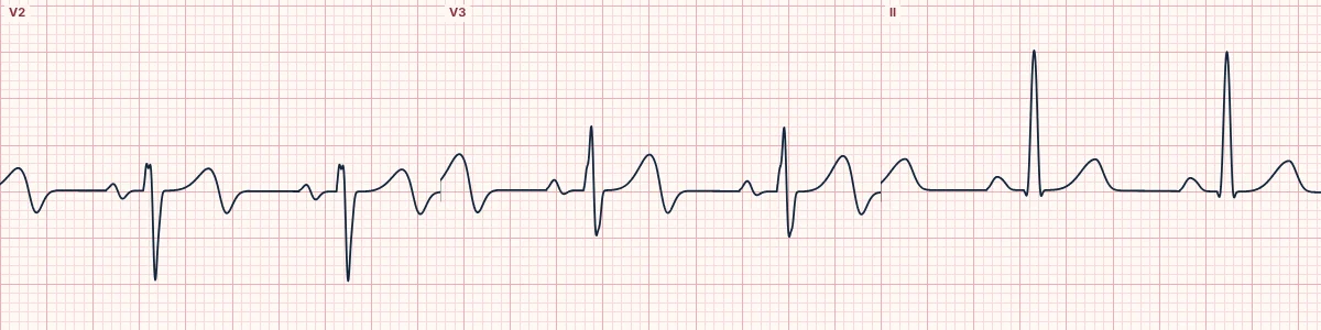
A warning sign of a critical narrowing in the proximal LAD, seen when the patient is pain-free: the T wave in V2–V3 goes up, then dips below the baseline.
What the heart is doing
After a brief LAD occlusion reopens, the anterior wall repolarizes abnormally. The last part of repolarization reverses direction, producing a terminal negative deflection on the T wave.
Why it looks this way
This is Wellens pattern type A because the T waves in V2–V3 are biphasic, positive then negative, with no significant ST elevation or Q waves.
Why it matters
Signals a high risk of a large anterior infarction within days to weeks. Treat as an acute coronary syndrome needing urgent angiography; stress testing is dangerous and should be avoided.
What to look for
- Check the end of the T wave in V2 and V3: it finishes with a sharp downward dip.
- R waves are intact and there is no significant ST elevation.
- The pattern appears between episodes of pain and may normalize during pain.
Frequently asked questions
What are the ECG criteria for wellens pattern, type A (biphasic T)?
T waves: biphasic (up then down) in V2–V3, sometimes V4. ST segment: isoelectric or < 1 mm elevated. QRS: no Q waves; R-wave progression preserved. Context: recent chest pain, now resolved.
How is wellens pattern, type A (biphasic T) identified on an ECG?
This is Wellens pattern type A because the T waves in V2–V3 are biphasic, positive then negative, with no significant ST elevation or Q waves.
What can wellens pattern, type A (biphasic T) be mistaken for?
Normal variant T waves: the terminal part is not sharply negative. Hypokalemia: a down-up T–U complex, the opposite sequence. Pulmonary embolism: T inversion in V1–V4 together with lead III.
What causes wellens pattern, type A (biphasic T)?
Common causes: critical proximal LAD stenosis; spontaneous reperfusion of an LAD occlusion; coronary artery spasm; and unstable plaque with intermittent occlusion.