
Ischemia & prior infarction
Draft — pending owner review
Aslanger pattern
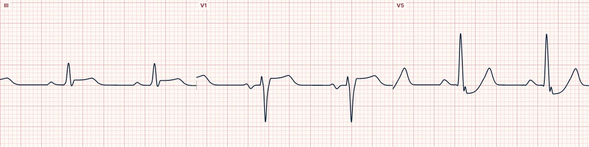
An inferior occlusion MI disguised by coexisting multivessel disease: ST elevation in lead III only, ST depression in V4–V6 and an ST segment higher in V1 than in V2.
What the heart is doing
A small inferior injury vector points down and to the right, and diffuse subendocardial ischemia adds a vector toward aVR. Their sum points rightward, so only III and aVR rise while the lateral chest leads are pulled down.
Why it looks this way
This is the Aslanger pattern because ST elevation is confined to lead III, V4–V6 show ST depression with upright T waves and the ST in V1 sits above V2.
Why it matters
These patients miss STEMI criteria and are often managed as non-ST-elevation ischemia, yet many have an occluded inferior artery. Recognize the pattern and push for emergent angiography; remember it can also persist after an old infarct.
What to look for
- Read lead III by itself: isolated ST elevation there is easy to dismiss.
- Check V4–V6 for ST depression with T waves that still point up.
- Compare the ST level in V1 with V2: higher in V1 supports the pattern.
Frequently asked questions
What are the ECG criteria for aslanger pattern?
Lead III: ST elevation, without significant elevation in II or aVF. V4–V6: ST depression with an upright or terminally upright T wave. V1 and V2: ST segment higher in V1 than in V2. aVR: often slight ST elevation.
How is aslanger pattern identified on an ECG?
This is the Aslanger pattern because ST elevation is confined to lead III, V4–V6 show ST depression with upright T waves and the ST in V1 sits above V2.
What can aslanger pattern be mistaken for?
Distal RCA occlusion: ST elevation in II, III and aVF. Subendocardial ischemia: widespread ST depression with no elevation in III. Left main coronary insufficiency: elevation in aVR and V1 with widespread depression.
What causes aslanger pattern?
Common causes: acute inferior occlusion (RCA or circumflex) with multivessel disease; diffuse subendocardial ischemia adding a vector toward aVR; and A prior infarction changing the overall ST vector.