
Ischemia and infarction
ST-elevation criteria
The thresholds and their limits
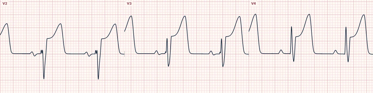
ST elevation at the J point in two contiguous leads, above lead-specific thresholds, defines ST-elevation myocardial infarction in the right clinical setting. The thresholds are higher in V2 and V3, where some elevation is normal.
The thresholds
New ST elevation at the J point in two contiguous leads of at least 1 mm in all leads other than V2–V3. In V2–V3: at least 2 mm in men aged 40 or over, 2.5 mm in men under 40 and 1.5 mm in women of any age. Posterior: at least 0.5 mm in V7–V9. Right ventricle: at least 0.5 mm in V3R–V4R.
Contiguous leads
Leads that look at neighbouring parts of the heart: II, III and aVF (inferior); I and aVL (high lateral); V1–V6 in sequence. Elevation in II and V2 alone is not contiguous.
Beyond the numbers
Some coronary occlusions do not reach the thresholds: hyperacute T waves, de Winter T waves, posterior infarction, left main patterns and occlusions in left bundle branch block or paced rhythms. Reciprocal depression and the shape of the ST segment often reveal occlusion before the millimetres do.
How to read it
- Measure at the J point against the TP baseline.
- Confirm two contiguous leads.
- Look for reciprocal depression and serial change; repeat the ECG if symptoms continue.
Pitfalls
- Left ventricular hypertrophy, bundle branch block, pericarditis, early repolarization, Brugada pattern and ventricular aneurysm cause ST elevation without acute occlusion.
Frequently asked questions
ST-elevation criteria: what is the key idea?
ST elevation at the J point in two contiguous leads, above lead-specific thresholds, defines ST-elevation myocardial infarction in the right clinical setting. The thresholds are higher in V2 and V3, where some elevation is normal.
ST-elevation criteria: what are the key numbers?
Most leads: ≥ 1 mm; V2–V3, men ≥ 40: ≥ 2 mm; V2–V3, men < 40: ≥ 2.5 mm; V2–V3, women: ≥ 1.5 mm; V7–V9: ≥ 0.5 mm; V3R–V4R: ≥ 0.5 mm.
ST-elevation criteria: how do you apply it to an ECG?
Measure at the J point against the TP baseline. Confirm two contiguous leads. Look for reciprocal depression and serial change; repeat the ECG if symptoms continue.
ST-elevation criteria: what are the common pitfalls?
Left ventricular hypertrophy, bundle branch block, pericarditis, early repolarization, Brugada pattern and ventricular aneurysm cause ST elevation without acute occlusion.