
Ischemia and infarction
Coronary territories and their leads
Which artery, which leads
Each group of leads looks at a wall of the left ventricle, and each wall is supplied by a predictable artery. Matching the leads to the territory points to the culprit vessel.
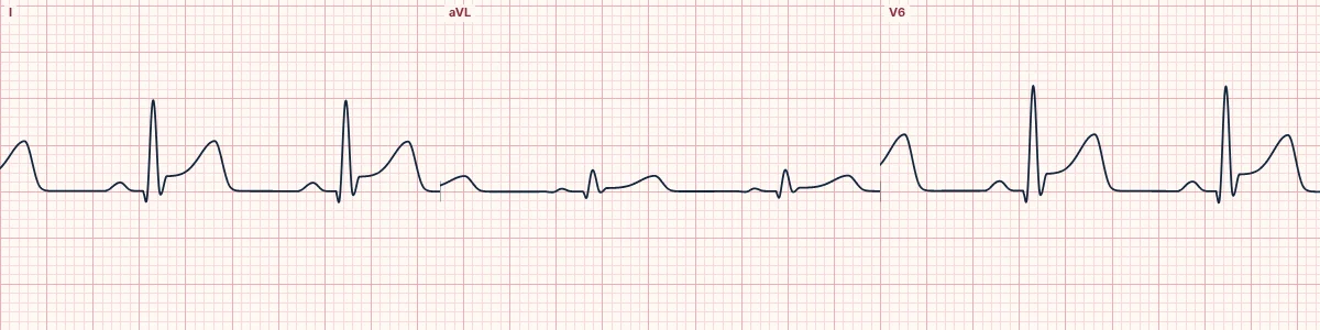
Anterior and septal
V1–V4 look at the septum and anterior wall, supplied by the left anterior descending artery. Proximal LAD occlusion often adds I and aVL (first diagonal) and can involve V5–V6.
Lateral
I, aVL, V5 and V6 look at the lateral wall, supplied by diagonal branches of the LAD and the circumflex.
Inferior
II, III and aVF look at the inferior wall, supplied by the right coronary artery in most people and by the circumflex in the rest. Elevation greater in III than II, with depression in I and aVL, favours the right coronary artery.
Posterior and right ventricle
The posterior wall (circumflex or right coronary) is seen in reverse in V1–V3 or directly in V7–V9. The right ventricle (proximal right coronary) is seen in V4R.
How to read it
- Group the abnormal leads by territory.
- Use the coronary panel in the lab to see the vessel and the muscle it supplies.
- Look for the reciprocal territory to confirm.
Pitfalls
- Coronary anatomy varies; dominance and wraparound LADs change which leads are involved.
Frequently asked questions
Coronary territories and their leads: what is the key idea?
Each group of leads looks at a wall of the left ventricle, and each wall is supplied by a predictable artery. Matching the leads to the territory points to the culprit vessel.
Coronary territories and their leads: what are the key numbers?
V1–V2: Septal: LAD; V3–V4: Anterior: LAD; I, aVL, V5–V6: Lateral: diagonals, circumflex; II, III, aVF: Inferior: RCA (or circumflex); V7–V9: Posterior: circumflex or RCA; V4R: Right ventricle: proximal RCA.
Coronary territories and their leads: how do you apply it to an ECG?
Group the abnormal leads by territory. Use the coronary panel in the lab to see the vessel and the muscle it supplies. Look for the reciprocal territory to confirm.
Coronary territories and their leads: what are the common pitfalls?
Coronary anatomy varies; dominance and wraparound LADs change which leads are involved.