
Position & lead placement
Dextrocardia
The heart lies on the right side of the chest as a mirror image. Standard lead placement then records the frontal plane reversed, with shrinking chest-lead complexes.
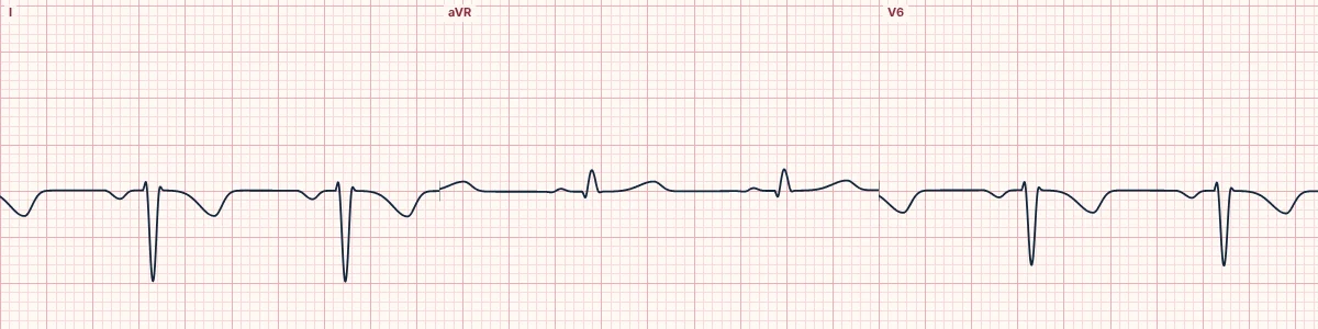
What the heart is doing
Activation spreads through a normal conduction system but from left to right across the chest. Lead I sees it moving away, and the left-sided chest electrodes sit far from the heart.
Why it looks this way
This is dextrocardia because lead I is globally inverted with an upright aVR, and the R wave shrinks progressively from V1 to V6.
Why it matters
Usually harmless when all organs are reversed. The practical point is to record a right-sided ECG for interpretation, and to place defibrillation pads to match the heart's position.
What to look for
- Everything in lead I is upside down, including the P wave.
- The chest leads get smaller toward V6 because they move away from the heart.
- Repeat with the chest leads on the right side and arm leads swapped: the ECG normalizes.
Frequently asked questions
What are the ECG criteria for dextrocardia?
Lead I: P wave, QRS and T wave all inverted. aVR: upright complexes (aVR and aVL appear swapped). Axis: right axis deviation. V1–V6: R wave shrinks from V1 to V6 (reversed progression).
How is dextrocardia identified on an ECG?
This is dextrocardia because lead I is globally inverted with an upright aVR, and the R wave shrinks progressively from V1 to V6.
What can dextrocardia be mistaken for?
Arm lead reversal: the same limb-lead picture but normal chest leads. Lateral infarction: Q waves in I and aVL with an upright P wave. Right ventricular hypertrophy: right axis with a tall R in V1.
What causes dextrocardia?
Common causes: congenital mirror-image position of the heart; complete reversal of the organs (situs inversus); primary ciliary dyskinesia; and associated congenital heart defects.