
Conduction system disease
Draft — pending owner review
Masquerading bundle branch block
RBBB in the chest leads, LBBB in the limb leads
An RBBB pattern in the chest leads with an LBBB look in the limb leads: no S wave in I and aVL and marked left axis deviation. It marks severe conduction system disease with a high risk of complete heart block.
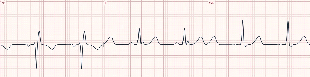
Standard form
V1 shows the rsR′ of RBBB, but lead I and aVL have a broad, slurred R wave with no S wave, so the limb leads look like LBBB. A severe left anterior fascicular block, often with left ventricular enlargement, swings the late rightward forces of the RBBB out of the limb leads.
Precordial form
RBBB shape in V1–V3, with an LBBB-like broad R wave and no S wave in V5–V6.
Why it matters
It reflects more extensive disease of the left bundle and the left ventricle than ordinary bifascicular block. Progression to complete AV block and mortality are high, so these patients need close follow-up and a pacing assessment.
About the demonstration
The lab draws ordinary bifascicular block (RBBB with left anterior fascicular block), which still has an S wave in I. Compare it with the description: in the masquerading form that S wave disappears.
How to read it
- When V1 says RBBB, check lead I for the S wave RBBB should produce.
- If it is missing, look at the axis and aVL.
- Treat it as advanced conduction disease.
Pitfalls
- It is easily filed as plain RBBB or plain LBBB depending on which leads are read first.
Frequently asked questions
Masquerading bundle branch block: what is the key idea?
An RBBB pattern in the chest leads with an LBBB look in the limb leads: no S wave in I and aVL and marked left axis deviation. It marks severe conduction system disease with a high risk of complete heart block.
Masquerading bundle branch block: what are the key numbers?
Key difference from bifascicular block: Absent S wave in I and aVL; Risk: High rate of progression to complete heart block; Associations: Ischemic and hypertensive heart disease, Lenègre–Lev disease.
Masquerading bundle branch block: how do you apply it to an ECG?
When V1 says RBBB, check lead I for the S wave RBBB should produce. If it is missing, look at the axis and aVL. Treat it as advanced conduction disease.
Masquerading bundle branch block: what are the common pitfalls?
It is easily filed as plain RBBB or plain LBBB depending on which leads are read first.