
Leads and axis
The chest leads and where they go
V1 to V6 in the horizontal plane
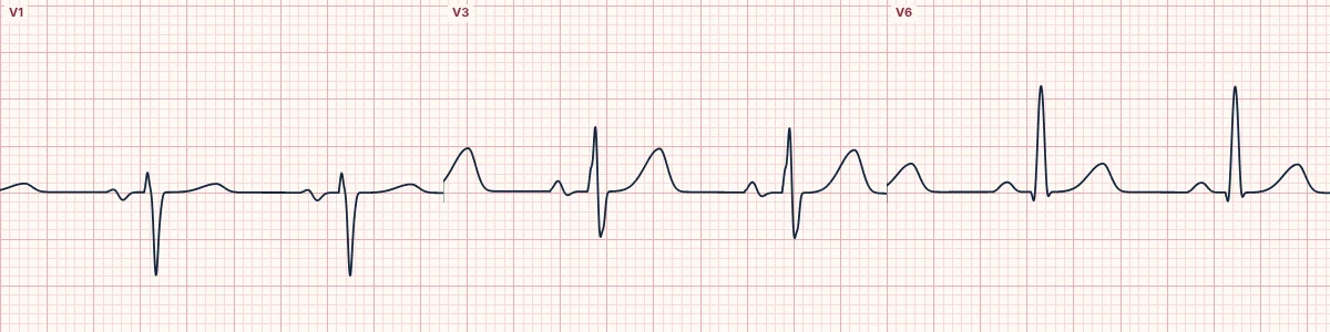
The six precordial leads sit on the chest wall in a horizontal line around the heart, from the right of the sternum (V1) to under the left arm (V6). Each one looks at the part of the heart closest to it.
Placement
V1 is in the fourth intercostal space at the right sternal edge and V2 in the fourth space at the left sternal edge. V4 is in the fifth space in the midclavicular line, and V3 halfway between V2 and V4. V5 is in the anterior axillary line and V6 in the midaxillary line, both level with V4.
What each one sees
V1 and V2 face the septum and the right ventricle, V3 and V4 the anterior wall, and V5 and V6 the lateral wall of the left ventricle. Because the electrodes are close to the heart, they are far more sensitive to local change than the limb leads.
Seeing the horizontal plane
Switch the heart panel to Chest leads 3D to see the electrodes around the heart and watch each one light up as the cardiac vector swings toward it.
How to read it
- Count the rib spaces from the angle of Louis (second rib) to place V1 and V2.
- Place V4 before V3.
- Keep V5 and V6 horizontal with V4, not following the curve of the rib.
Pitfalls
- Placing V1 and V2 one space too high produces rSr′ patterns and inverted P waves that mimic disease.
- Placing chest leads under rather than on the breast changes amplitudes; place them on the chest wall beneath the breast tissue if needed, at the correct level.
Frequently asked questions
The chest leads and where they go: what is the key idea?
The six precordial leads sit on the chest wall in a horizontal line around the heart, from the right of the sternum (V1) to under the left arm (V6). Each one looks at the part of the heart closest to it.
The chest leads and where they go: what are the key numbers?
V1: 4th ICS, right sternal edge; V2: 4th ICS, left sternal edge; V3: Midway V2–V4; V4: 5th ICS, midclavicular line; V5: Anterior axillary line, level of V4; V6: Midaxillary line, level of V4.
The chest leads and where they go: how do you apply it to an ECG?
Count the rib spaces from the angle of Louis (second rib) to place V1 and V2. Place V4 before V3. Keep V5 and V6 horizontal with V4, not following the curve of the rib.
The chest leads and where they go: what are the common pitfalls?
Placing V1 and V2 one space too high produces rSr′ patterns and inverted P waves that mimic disease. Placing chest leads under rather than on the breast changes amplitudes; place them on the chest wall beneath the breast tissue if needed, at the correct level.