
Special populations
Draft — pending owner review
Pediatric ECG basics
Normal values change with age
A child's ECG is read against values for the child's age. Faster rates, a rightward axis, right ventricular dominance and inverted T waves in V1–V3 are all normal in infants and young children.
Why children are different
Before birth the right ventricle pumps against the high resistance of the unexpanded lungs, so at birth it is as thick as the left. Its forces dominate the newborn ECG. Over the first years the left ventricle grows to dominate, and the ECG moves toward the adult pattern by adolescence.
Right ventricular dominance
Infants have a rightward axis and tall R waves in V1, often with deep S waves in V6. In an adult these would mean right ventricular hypertrophy; in a baby they are expected. Judge them against age-specific normal tables rather than adult criteria.
The juvenile T-wave pattern
T waves in V1 are upright for the first few days of life, then invert by about one week and usually stay inverted in V1–V3 until adolescence, sometimes longer. An upright T wave in V1 between about one week and adolescence is a recognized sign of right ventricular hypertrophy.
Rates and intervals
Resting rates are faster the younger the child, and the PR interval and QRS duration are shorter. A QRS that would be narrow for an adult can be wide for an infant. The QTc upper limit is similar to adults, slightly higher in the first months of life.
About the demonstration
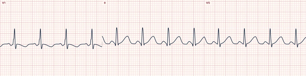
The live tracing shows a simulated infant pattern: sinus rhythm at 125/min with a rightward axis, a dominant R wave in V1 and inverted T waves in V1–V3. It is an illustration of the main features, not a reference for measuring a real child.
How to read it
- Find the age before anything else.
- Read rate, axis, intervals and V1 against age-specific normal values.
- Check T waves in V1: inverted is normal in childhood; upright after the first week deserves a second look.
Pitfalls
- Applying adult voltage criteria over-calls hypertrophy in children, whose chests are thin and close to the electrodes.
- Sinus arrhythmia is marked in children and is normal.
- Electrode placement on a small chest crowds the leads; V1 and V2 placed too high change the pattern.
Frequently asked questions
Pediatric ECG basics: what is the key idea?
A child's ECG is read against values for the child's age. Faster rates, a rightward axis, right ventricular dominance and inverted T waves in V1–V3 are all normal in infants and young children.
Pediatric ECG basics: what are the key numbers?
Newborn: Rate about 90–160/min; axis about +30° to +180°; 1–12 months: Rate about 100–160/min; axis about +10° to +120°; 1–5 years: Rate about 80–140/min; axis about 0° to +110°; 6–12 years: Rate about 65–120/min; axis about −15° to +110°; Adolescent: Close to adult values; T in V1: Upright in the first days; inverted from about 1 week until adolescence; Values: Approximate: use a validated age-specific table.
Pediatric ECG basics: how do you apply it to an ECG?
Find the age before anything else. Read rate, axis, intervals and V1 against age-specific normal values. Check T waves in V1: inverted is normal in childhood; upright after the first week deserves a second look.
Pediatric ECG basics: what are the common pitfalls?
Applying adult voltage criteria over-calls hypertrophy in children, whose chests are thin and close to the electrodes. Sinus arrhythmia is marked in children and is normal. Electrode placement on a small chest crowds the leads; V1 and V2 placed too high change the pattern.