
Paper and measurement
The intervals: PR, QRS and QT
What normal looks like
Three intervals describe how long each part of the beat takes: the PR (atria to ventricles), the QRS (ventricular activation) and the QT (ventricular activation and recovery).
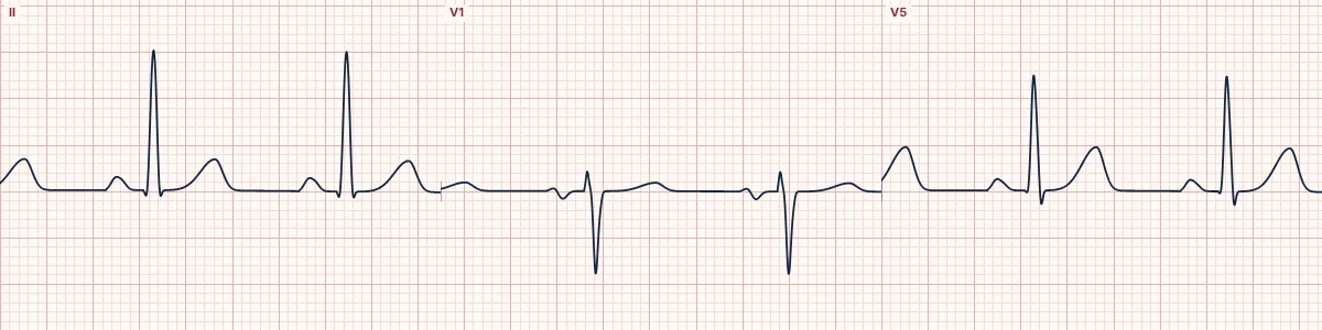
PR interval
From the start of the P wave to the start of the QRS. It contains atrial activation and the deliberate pause in the AV node. Normal is 120–200 ms. Longer means slow AV conduction; shorter suggests a pathway that bypasses the AV node or a junctional origin.
QRS duration
From the first deflection of the QRS to the J point. A normal QRS is under 120 ms (usually 80–100 ms) because the His–Purkinje system activates the ventricles almost at once. A wide QRS means the impulse is travelling muscle to muscle: bundle branch block, a ventricular origin, pre-excitation, pacing or drugs and electrolytes that slow conduction.
QT interval
From the start of the QRS to the end of the T wave. It shortens as the heart rate rises, so it is corrected for rate (QTc) before being judged. Measure it in the lead with the longest QT, usually II or V5, and exclude U waves.
How to read it
- Measure each interval in the lead where it is longest and its edges clearest.
- Pause the live tracing and use Calipers to measure PR, QRS and QT on a real beat.
- Correct the QT for rate before calling it long or short.
Pitfalls
- A wide QRS also lengthens the QT; judge repolarization separately (for example, JT interval) in bundle branch block.
- A U wave merging with the T wave makes the QT look falsely long.
Frequently asked questions
The intervals: PR, QRS and QT: what is the key idea?
Three intervals describe how long each part of the beat takes: the PR (atria to ventricles), the QRS (ventricular activation) and the QT (ventricular activation and recovery).
The intervals: PR, QRS and QT: what are the key numbers?
PR: 120–200 ms (3–5 small boxes); QRS: < 120 ms (< 3 small boxes); QTc, men: Prolonged above 450 ms; QTc, women: Prolonged above 460–470 ms; QTc: > 500 ms: high risk of torsades.
The intervals: PR, QRS and QT: how do you apply it to an ECG?
Measure each interval in the lead where it is longest and its edges clearest. Pause the live tracing and use Calipers to measure PR, QRS and QT on a real beat. Correct the QT for rate before calling it long or short.
The intervals: PR, QRS and QT: what are the common pitfalls?
A wide QRS also lengthens the QT; judge repolarization separately (for example, JT interval) in bundle branch block. A U wave merging with the T wave makes the QT look falsely long.