
Heart muscle and systemic disease
Draft — pending owner review
Restrictive and infiltrative cardiomyopathy
Thick walls, small complexes
In amyloidosis and other infiltrative diseases the walls look thick on imaging but the QRS voltage is low. Low voltage, a pseudo-infarct pattern and conduction disease together should prompt the diagnosis.
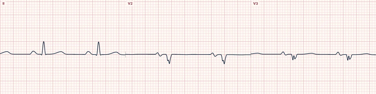
The voltage mismatch
Hypertrophy of muscle raises voltage. Thickening from deposits that do not conduct (amyloid, granulomas, iron) does not, and it insulates the surviving muscle. Low limb-lead voltage with thick walls on echocardiography is a strong clue to cardiac amyloidosis.
Other findings
Poor R-wave progression and Q waves without an infarct (a pseudo-infarct pattern), atrial fibrillation, AV block and bundle branch block. Sarcoidosis characteristically causes AV block and VT in younger patients.
Why it matters
Treatable forms exist, so an unexplained mismatch between thick walls and small complexes should not be put down to body habitus.
About the demonstration
The tracing combines low QRS voltage with loss of anterior R waves to show the low-voltage, pseudo-infarct picture.
How to read it
- Check limb-lead voltage against any report of wall thickness.
- Look for Q waves without a matching history of infarction.
- Note conduction disease and atrial fibrillation.
Pitfalls
- Obesity, effusion and lung disease also lower voltage.
- Some patients with amyloidosis still meet LVH voltage criteria.
Frequently asked questions
Restrictive and infiltrative cardiomyopathy: what is the key idea?
In amyloidosis and other infiltrative diseases the walls look thick on imaging but the QRS voltage is low. Low voltage, a pseudo-infarct pattern and conduction disease together should prompt the diagnosis.
Restrictive and infiltrative cardiomyopathy: what are the key numbers?
Low voltage: QRS < 5 mm in every limb lead; Pseudo-infarct: Q waves or poor R progression without infarction; Conduction: AV block and bundle branch block are common.
Restrictive and infiltrative cardiomyopathy: how do you apply it to an ECG?
Check limb-lead voltage against any report of wall thickness. Look for Q waves without a matching history of infarction. Note conduction disease and atrial fibrillation.
Restrictive and infiltrative cardiomyopathy: what are the common pitfalls?
Obesity, effusion and lung disease also lower voltage. Some patients with amyloidosis still meet LVH voltage criteria.