
The waves
Pathological Q waves
The scar of an old infarction
Dead muscle produces no electrical activity, so leads over a scar look straight through it and record the opposite wall moving away: a wide or deep Q wave.
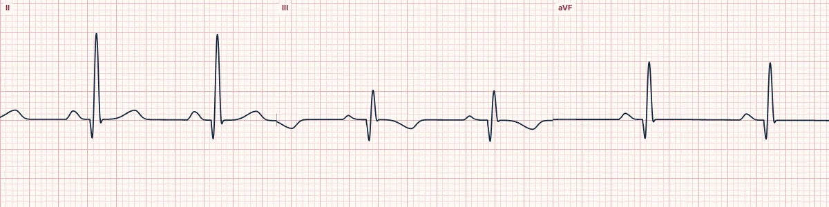
When a Q wave is abnormal
Any Q wave in V2 or V3 lasting 20 ms or more (or a QS complex), or a Q wave of 30 ms or more and at least 1 mm deep in two contiguous leads elsewhere, is consistent with prior infarction. An older rule of thumb is a Q wave over 40 ms wide or more than a quarter of the R wave's height.
Normal Q waves
Small septal q waves in I, aVL, V5 and V6 are normal. An isolated Q wave in III that disappears with deep inspiration is usually positional.
Other causes
Hypertrophic cardiomyopathy (narrow, deep septal Q waves), pre-excitation, left bundle branch block, lead misplacement and infiltrative disease can all produce Q waves without infarction.
How to read it
- Group Q waves by territory.
- Compare with old ECGs to date them.
- Persistent ST elevation with Q waves weeks after an infarct suggests a ventricular aneurysm.
Pitfalls
- A QS in V1 alone is common and normal.
Frequently asked questions
Pathological Q waves: what is the key idea?
Dead muscle produces no electrical activity, so leads over a scar look straight through it and record the opposite wall moving away: a wide or deep Q wave.
Pathological Q waves: what are the key numbers?
V2–V3: Any Q ≥ 20 ms, or QS; Other leads: Q ≥ 30 ms and ≥ 1 mm, in 2 contiguous leads; Rule of thumb: > 40 ms or > 25% of R.
Pathological Q waves: how do you apply it to an ECG?
Group Q waves by territory. Compare with old ECGs to date them. Persistent ST elevation with Q waves weeks after an infarct suggests a ventricular aneurysm.
Pathological Q waves: what are the common pitfalls?
A QS in V1 alone is common and normal.