
The waves
The P wave
Atrial depolarization
The P wave is the right and left atria depolarizing one after the other. Its shape tells you where the impulse started and whether the atria are enlarged.
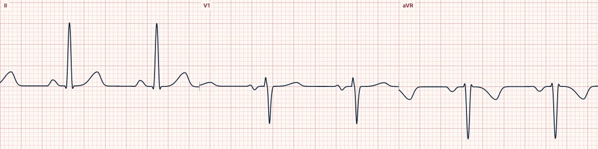
Normal P
Smooth and rounded, under 120 ms long and under 2.5 mm tall in lead II. Upright in I, II and aVF, inverted in aVR, and often biphasic in V1 because the right atrium (in front) activates before the left atrium (behind).
Where did it start?
A sinus P wave travels down and to the left, so its axis is roughly 0° to +75°. An inverted P in II, III and aVF means the atria are being activated from below: a low atrial or junctional origin.
Enlarged atria
A tall, peaked P (≥ 2.5 mm in II) suggests right atrial enlargement. A long, notched P (≥ 120 ms) with a deep negative terminal part in V1 suggests left atrial enlargement.
How to read it
- Find a P wave before every QRS and a QRS after every P.
- Check that P is upright in II and inverted in aVR.
- Look at V1 for the biphasic shape and the size of its negative part.
Pitfalls
- P waves can hide in the T wave of the beat before; look for a T wave that is shaped differently from its neighbours.
Frequently asked questions
The P wave: what is the key idea?
The P wave is the right and left atria depolarizing one after the other. Its shape tells you where the impulse started and whether the atria are enlarged.
The P wave: what are the key numbers?
Duration: < 120 ms; Height in II: < 2.5 mm; Axis: 0° to +75°; aVR: Inverted.
The P wave: how do you apply it to an ECG?
Find a P wave before every QRS and a QRS after every P. Check that P is upright in II and inverted in aVR. Look at V1 for the biphasic shape and the size of its negative part.
The P wave: what are the common pitfalls?
P waves can hide in the T wave of the beat before; look for a T wave that is shaped differently from its neighbours.